Superior vestibular area (Latin: area vestibularis superior)
The superior vestibular area is one of the four areas into which the fundus of the internal acoustic meatus is divided; it lies in the back of the upper part.
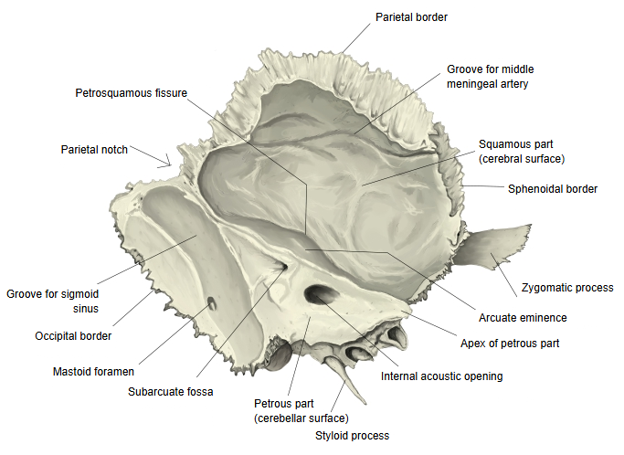
Fundus of the internal acoustic meatus: the blind end of the internal acoustic meatus, which starts at the internal acoustic openingThe internal acoustic opening lies on the posterior surface of the petrous part of the temporal bone
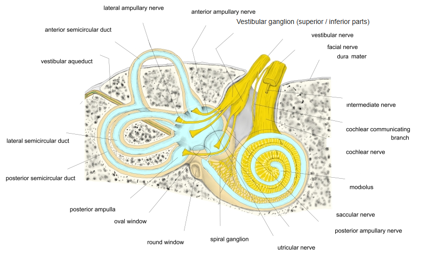
In the superior vestibular area open many small holes; they lead to the superior cribriform area of the vestibule and transmit the utriculo-ampullary nerve.
Superior cribriform area: in the elliptical recess, above the pyramid of the vestibuleUtriculo-ampullary nerve: the upper part of the vestibular ganglion continues into it; it then divides into three branches
Fundus of the
internal
acoustic
meatus
internal
acoustic
meatus
- Upper part
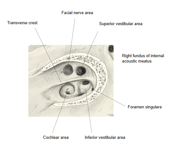
- Front: Area of facial nerve : the opening of the facial canal, through which the facial nerve passes.
- Back: Superior vestibular area : many small holes leading to the superior cribriform area of the vestibule; the utriculo-ampullary nerve passes through them.
- Lower part
- Front: Cochlear area : many small holes arranged in a spiral, through which the cochlear nerve passes.
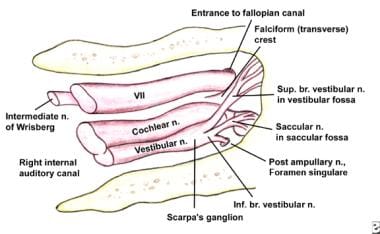
- Back: Inferior vestibular area : small holes leading to the middle cribriform area of the vestibule, for the saccular nerve. Behind and below it lies a single hole, the foramen singulare, which leads to the inferior cribriform area and transmits the posterior ampullary nerve.
· The transverse crest divides the fundus into upper and lower parts, each with a front and a back area. The superior vestibular area lies above the crest and behind the area of facial nerve.
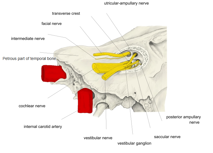
· The utriculo-ampullary nerve that passes through it comes from the upper part of the vestibular ganglion and divides into the utricular, anterior ampullary and lateral ampullary nerves.
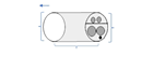 Internal acoustic meatus (diagram)
 Areas of the fundus (diagram)
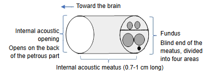
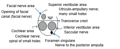
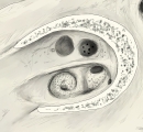 Fundus of the right internal acoustic meatus
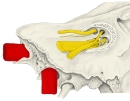 Nerves at the fundus
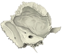 Left temporal bone (medial surface)
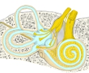 Inner ear and its nerves
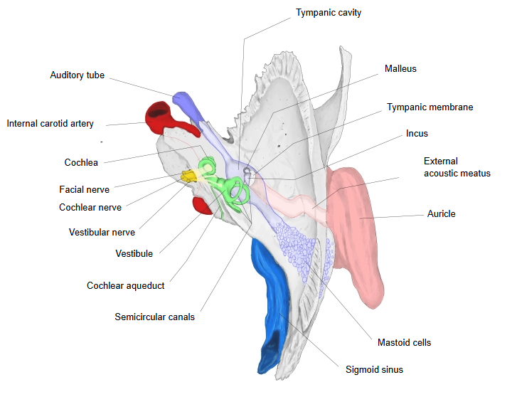
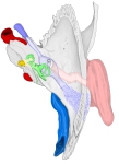 Right temporal bone and ear (superior view)
Reference websites
[Wikipedia]
· Internal auditory meatus, Wikipedia.
· Vestibular nerve, Wikipedia.
[Websites with illustrations]
· The four areas of the fundus of the internal acoustic meatus (diagram) (Folia Morphologica)
· Nerves in the right internal auditory canal (drawing) (Medscape)
Related terms
- Superior cribriform area
- Fundus of internal acoustic meatus
- Internal acoustic meatus
- Transverse crest
- Area of facial nerve
- Cochlear area
- Vestibular ganglion
- Utriculo ampullar nerve
References and sources
- Further reading: Internal auditory meatus, Wikipedia.
- Further reading: Vestibular nerve, Wikipedia.
- Federative International Programme for Anatomical Terminology (FIPAT). Terminologia Anatomica, 2nd ed. (TA2), 2019.