Macula densa (Latin: macula densa)
Overview
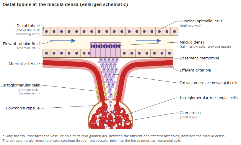
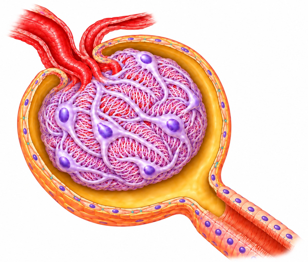
· The macula densa lies between the afferent arteriole and the efferent arteriole, in close contact with the juxtaglomerular cells in the wall of the afferent arteriole and with the extraglomerular mesangial cells.
· Its cells sense the sodium chloride (NaCl) concentration of the fluid flowing through the renal tubule. When the concentration falls, the afferent arteriole widens, the pressure in the glomerulus rises and the juxtaglomerular cells release more renin. When it rises, the afferent arteriole narrows and the glomerular filtration rate falls. This adjustment of the afferent arteriole is called tubuloglomerular feedback; the change in renin release is a separate response.
· Together with the juxtaglomerular cells and the extraglomerular mesangial cells, the macula densa forms the juxtaglomerular apparatus, the starting point of the renin-angiotensin-aldosterone system that regulates blood pressure and body fluid volume.
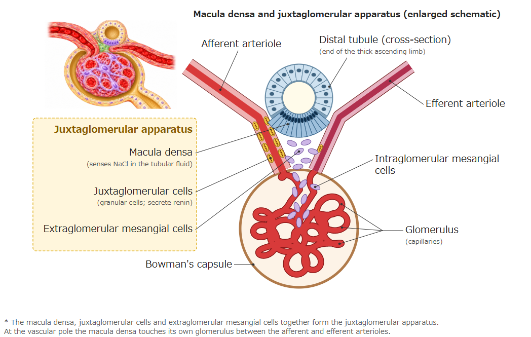
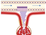 Macula densa (schematic)
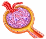 Renal corpuscle (section)
Reference websites
[Wikipedia]
· Macula densa, Wikipedia.
· Juxtaglomerular apparatus, Wikipedia.
· Tubuloglomerular feedback, Wikipedia.
[Websites with illustrations]
· Tubules and collecting system (8) (Digital Histology)
· Macula densa (micrograph) (SIU School of Medicine histology)
· Juxtaglomerular apparatus (micrograph) (SIU School of Medicine histology)
· Kidney histology (Tufts University histology images)
Related terms
References and sources
- Further reading: Macula densa, Wikipedia.
- Further reading: Juxtaglomerular apparatus, Wikipedia.
- Further reading: Tubuloglomerular feedback, Wikipedia.
- Federative International Programme for Anatomical Terminology (FIPAT). Terminologia Anatomica, 2nd ed. (TA2), 2019.