Pharyngeal arch (Latin: arcus pharyngeus)
Also called: branchial arch; visceral arch
= branchial arch; pharyngeal arch is the standard name, branchial arch the older one.
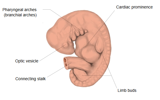
Pharyngeal arches are the paired, bar-like swellings on each side of the head and neck region of the early embryo, found in all vertebrate embryos; in humans they appear during the fourth and fifth weeks of development. Each arch has a core of mesenchyme with its own cartilage, muscle, artery and cranial nerve. Grooves (pharyngeal clefts) separate the arches on the outside, and pouches separate them on the inside. Six pairs are usually counted, but the fifth arch is absent or only rudimentary and forms no cartilage. They develop into many structures of the face, neck, pharynx and larynx: for example, the first arch gives the malleus and incus (and its cartilage is linked with the development of the mandible), the second the stapes and part of the hyoid bone, and the fourth and sixth arches the laryngeal cartilages such as the thyroid cartilage. The name branchial arch comes from fishes, where the arches behind the jaws support the gills; because the first arch forms the jaws and the second (hyoid) arch carries at most a half-gill in some fishes, some authors use the name only for the third and later arches. Sources differ.
Related terms
References and sources
- Further reading: Pharyngeal arch, Wikipedia.
- Federative International Programme for Anatomical Terminology (FIPAT). Terminologia Anatomica, 2nd ed. (TA2), 2019.