Pharynx (Latin: pharynx)
- Overview
- Parts (nasal part, oral part, laryngeal part)
- Communications
- Structure (mucosa, muscle layer, adventitia)
- Vessels and nerves
- Related terms
- References
Overview
The chart below shows, in simple form, the spaces that make up the respiratory and digestive systems.
* Accessory organs are left out.
system
- Airway
- Nasal cavity : from the nostrils to the choanae; divided into right and left halves by the nasal septum and lined with mucosa.
- Pharynx : made of three parts (upper, middle, lower) and lined with mucosa; also part of the digestive system.
- Larynx : funnel-shaped tube at the level of the 4th–7th cervical vertebrae; its framework is cartilage.
- Trachea : tube about 10 cm long and 20–25 mm wide; divides at the level of the 4th–5th thoracic vertebrae.
- Bronchi : branch from the trachea to the right and left, and divide into many bronchial branches inside the lungs.
- Lungs : right lung (upper, middle, lower lobes), left lung (upper, lower lobes); gas exchange (external respiration) takes place in the alveoli.
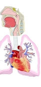
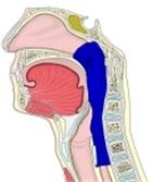 Pharynx
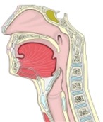 Nasal cavity to pharynx
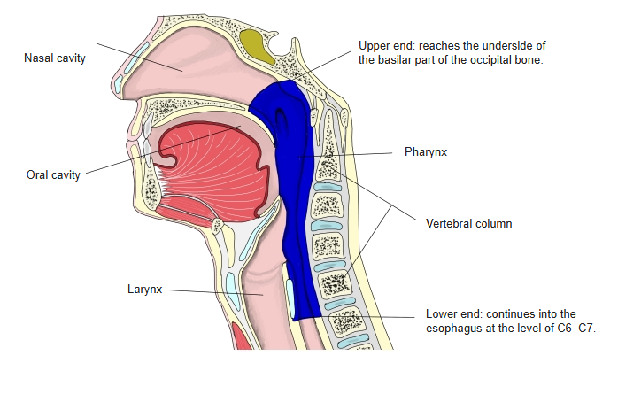
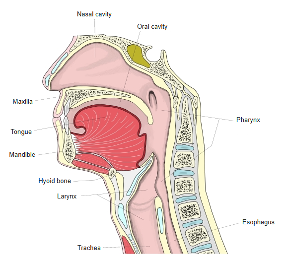
The upper end of the pharynx reaches the underside of the base of the occipital bone, and at the level of the 6th–7th cervical vertebrae it continues into the esophagus. It is about 12 cm long and shaped like a cone, wide above and narrow below.
Lower down it becomes more and more tube-like; its front and back walls come close together, and at the level of the larynx they touch, leaving only a slit, except while a bolus of food is passing into the esophagus.
The inside of the pharynx is lined with mucosa: in the nasal part it is pseudostratified ciliated columnar epithelium, and in the oral part and the laryngeal part it is stratified squamous epithelium.
Parts
The pharynx can be divided into the following three parts. See the page of each part for details.
| 1 | Nasal part of pharynx (= upper pharynx) | pars nasalis pharyngis (nasopharynx, epipharynx) | 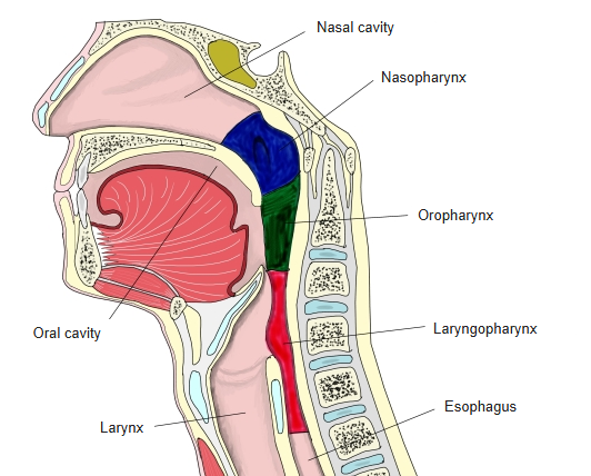 |
| 2 | Oral part of pharynx (= middle pharynx) | pars oralis pharyngis (oropharynx, mesopharynx) | |
| 3 | Laryngeal part of pharynx (= lower pharynx) | pars laryngea pharyngis (laryngopharynx, hypopharynx) |
the pharynx
- Nasopharynx (= nasal part) : opens into the nasal cavity through the choanae, and into the middle ear (auditory tube) through the pharyngeal opening of the auditory tube.
Related terms: torus levatorius, salpingopharyngeal fold, pharyngeal recess, torus tubarius, pharyngeal fornix, pharyngeal tonsil - Oropharynx (= oral part) : opens into the oral cavity through the isthmus of the fauces.
- Laryngopharynx (= laryngeal part) : opens into the larynx through the laryngeal inlet, and continues into the esophagus at the opening of the esophagus.
Related term: piriform recess
Communications
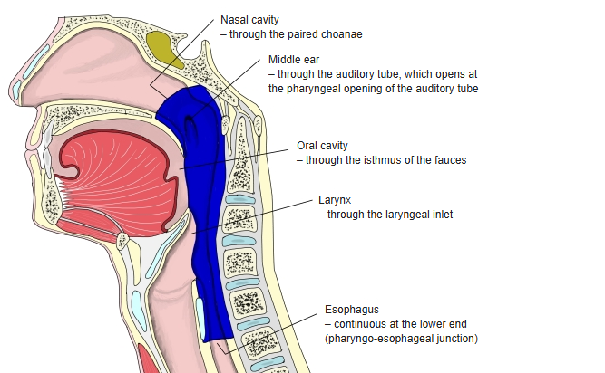
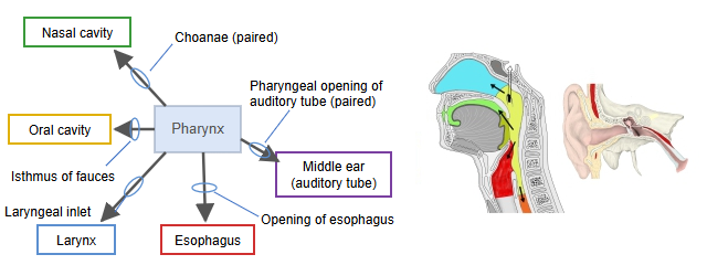
Through its openings the pharynx communicates with the following:
· Nasal cavity: through the choanae (paired).
· Oral cavity: through the isthmus of the fauces.
· Larynx: through the laryngeal inlet.
· Esophagus: through the opening of the esophagus.
· Middle ear: through the pharyngeal opening of the auditory tube (paired), by way of the auditory tube, with the middle ear (tympanic cavity).
Structure
■ Mucosa ■
The mucosa of the pharynx is pale pink.
| 1 | Front part of the nasopharynx | Made of pseudostratified ciliated columnar epithelium | ⇒ like the respiratory system |
| 2 | Back wall of the nasopharynx, the oropharynx and the laryngopharynx | Made of stratified squamous epithelium | ⇒ like the digestive system |
The lamina propria has no papillae where the ciliated epithelium lies, but forms papillae under the stratified squamous epithelium.
In the pharyngeal fornix, many lymphoid nodules gather to form the pharyngeal tonsil.
The pharyngeal glands are small, mainly mucous mixed glands: mixed in the fornix, more purely mucous lower down, and mixed again near the opening of the esophagus.
The pharyngeal mucosa has no muscularis mucosae yet.
Near the cranial base, where the muscle layer is missing, the submucosa is a tough fibrous membrane.
■ Muscle layer ■
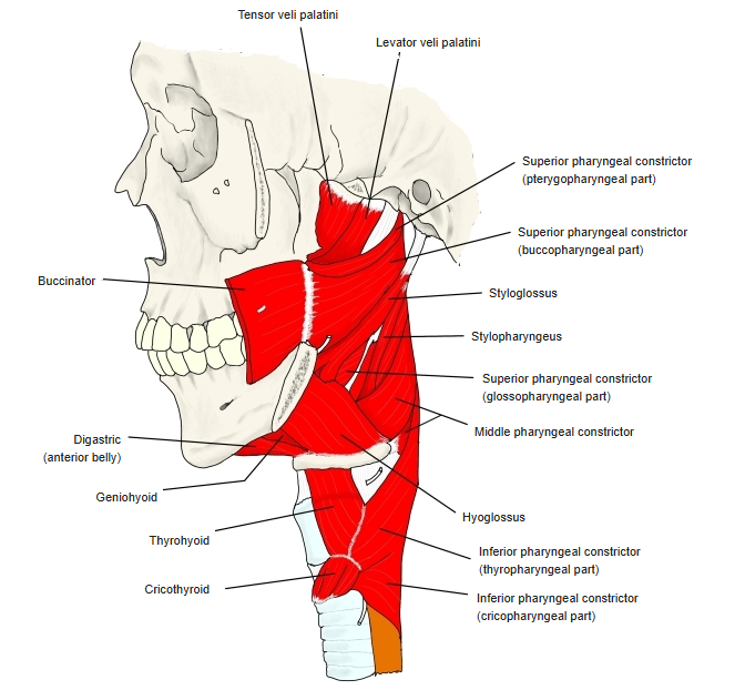
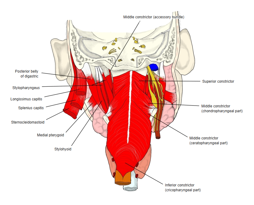
The muscle layer of the pharynx (striated muscle) consists of the following two groups.
of pharynx
- Longitudinal muscles : raise the pharynx. Pharyngeal elevators
- Circular muscles : constrict the pharynx. Pharyngeal constrictors
· At the very top, where there is no muscle, lies the pharyngobasilar fascia.
The chart below summarizes the muscles of the pharynx.
the pharynx
- Pharyngeal constrictors (circular)
- Superior constrictor : origin) medial pterygoid plate and pterygoid hamulus, pterygomandibular raphe, back end of the mylohyoid line of the mandible, side of the tongue.
- Middle constrictor : origin) greater and lesser horns of the hyoid bone and the lower end of the stylohyoid ligament.
- Inferior constrictor : origin) thyroid cartilage, cricoid cartilage.
- All three insert) pharyngeal raphe.
- Pharyngeal elevators (longitudinal)
- Palatopharyngeus : origin) palatine aponeurosis and posterior border of the hard palate (some fibres from the cartilage of the auditory tube); insertion) back border of the thyroid cartilage and pharyngeal wall.
- Salpingopharyngeus : origin) cartilage of the auditory tube; ends by joining the palatopharyngeus.
- Stylopharyngeus : origin) styloid process of the temporal bone; insertion) upper and back borders of the thyroid cartilage, side of the epiglottis and pharyngeal wall.
■ Adventitia ■
Made of loose connective tissue rich in elastic fibres.
Vessels and nerves
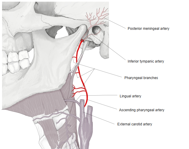
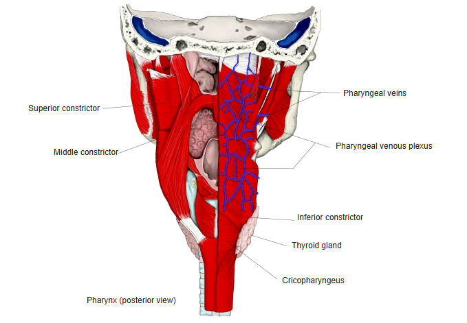
The vessels and nerves of the pharynx are as follows.
| 1 | Arteries | Ascending pharyngeal artery (from the external carotid artery) and branches of nearby arteries (facial, maxillary, lingual, superior and inferior thyroid) |
| 2 | Veins | Pharyngeal venous plexus (drains through the pharyngeal veins into the internal jugular vein) |
| 3 | Lymphatics | Upper part: to the retropharyngeal lymph nodes. Lower part: to the deep cervical lymph nodes. |
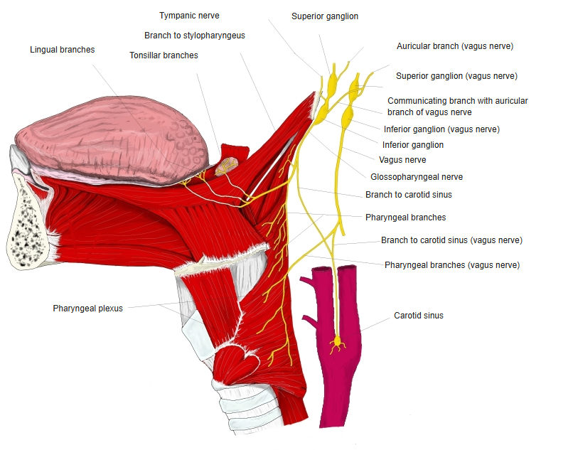
| 4 | Nerves | Pharyngeal constrictors: branches of the pharyngeal plexus, formed by the pharyngeal branches of the vagus, glossopharyngeal and sympathetic nerves. Pharyngeal elevators: the stylopharyngeus is supplied by the glossopharyngeal nerve; the others by the pharyngeal plexus. |
Related terms
Main anatomical terms related to the pharynx, in alphabetical order.
References and sources
- Further reading: Pharynx, Wikipedia.