Synapse (Latin: synapsis)
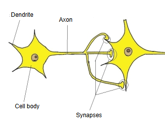
A typical synapse lies between an axon terminal of one neuron, the presynaptic side, and the dendrite, cell body or axon of the next cell, the postsynaptic side.
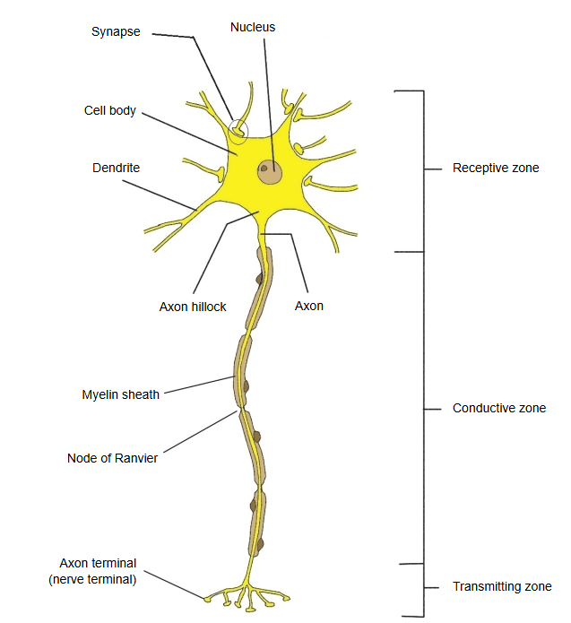
Along a neuron, the dendrites and cell body form the receiving zone, the axon the conducting zone, and the axon terminals with their synapses the transmitting zone.
- Chemical synapse
(vesicular synapse) : a neurotransmitter is released in one direction (presynaptic cell ⇒ postsynaptic cell) between the cells that pass the signal. Examples: between neurons; from a neuron to a muscle. - Electrical synapse
(non-vesicular synapse) : the cells are joined by tiny channels called connexons, and ions pass through them, so the signal can travel in both directions. Examples: neurons of the retina, heart muscle. - Mixed synapse : chemical and electrical transmission occur together at the same junction.
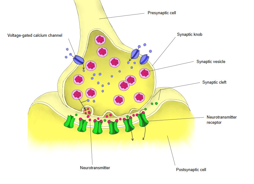
Chemical synapse: the terminal releases neurotransmitter from synaptic vesicles into a narrow synaptic cleft (about 20 nm wide); the transmitter binds to receptors on the postsynaptic membrane. Transmission is one-way and has a short delay. It is the usual type between neurons and at the junctions between nerve and muscle.
Electrical synapse: the two cell membranes are joined by a gap junction (a gap of about 3.5 nm; sources differ slightly) made of connexons. Ions pass directly from cell to cell, so transmission is very fast and can run both ways.
[Parts of a synapse]
· Presynaptic part: the swollen end of the axon (synaptic knob or bouton), with synaptic vesicles and voltage-gated calcium channels; calcium entry triggers the release of transmitter.
· Synaptic cleft: the narrow space between the two cells.
· Postsynaptic part: the membrane of the receiving cell, with receptors for the transmitter.
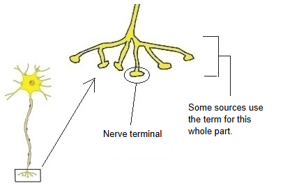
Some texts use "synapse" or "nerve terminal" for the whole terminal branching of an axon, others only for the single point of contact.
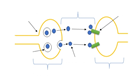 Chemical synapse (schematic)
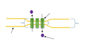 Electrical synapse (schematic)
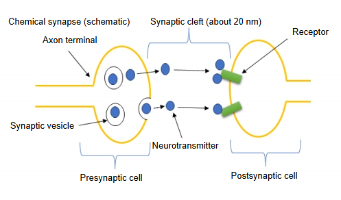
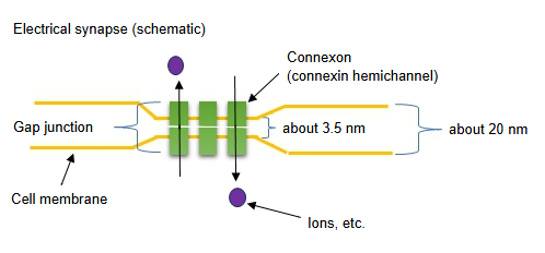
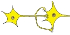 Synapses between two neurons
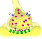 Structure of a chemical synapse
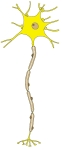 Neuron
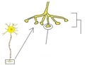 Nerve terminal
Reference websites
[Wikipedia]
· Synapse, Wikipedia.
· Gap junction, Wikipedia.
[Websites with illustrations]
· Schematic of a chemical synapse (Wikimedia Commons)

· Synaptic transmission (illustration) (Nuronlabs)

· The synapse (slide) (SlideShare)

Related terms
- Axon
- Dendrite
- Chemical synapse
- Electrical synapse
- Axon terminal
- Interneuron
- Nervous tissue
- Node of Ranvier
References and sources
- Further reading: Synapse, Wikipedia.
- Further reading: Gap junction, Wikipedia.
- Federative International Programme for Anatomical Terminology (FIPAT). Terminologia Anatomica, 2nd ed. (TA2), 2019.