Rod cell
Also called Rod or the rod photoreceptor
receptor
cells
- Rod cell : rod-shaped outer segment; the great majority of photoreceptors. Very sensitive, it serves light reception in dim light, without colour.
- Cone cell : cone-shaped outer segment; concentrated in the centre of the retina (macula and fovea). It works in bright light and gives sharp and colour vision.
of the
optic part
of the
retina
- Photoreceptor cells
- Rod cell : rod-shaped outer segment; most photoreceptors are rods. They serve light reception in dim light.
- Cone cell : cone-shaped outer segment; concentrated in the centre of the retina. They work in bright light and give sharp and colour vision.
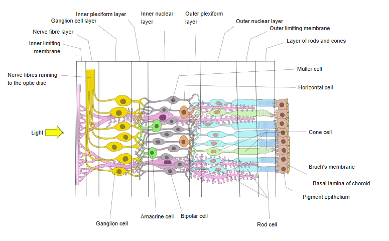
- Horizontal cell : its processes spread sideways along the retina and synapse with the photoreceptor cells.
- Bipolar cell : has two processes, one on each side of the cell body; passes the signal from the photoreceptors to the ganglion cells.
- Amacrine cell : a local interneuron without a long axon; links bipolar cells and ganglion cells.
- Ganglion cell : has a long axon that runs to the brain; the axons form the optic nerve, optic chiasm and optic tract.
- Müller cell : not a neuron but a kind of glial cell; it spans the retina as a supporting cell and does not carry the visual signal.
· In the periphery of the retina rods far outnumber cones, but towards the macula the relation is reversed and rods become gradually fewer; the centre of the fovea has none.
· The human retina has roughly 90-120 million rods against about 6 million cones (sources differ).
· Rods are a little longer and slimmer than cones, but both have the same basic structure.
· A human rod cell is about 2 µm thick; given lengths range from about 60 to 100 µm.
Rods lie in the outermost layer of the neural retina (the layer of rods and cones), just inside the retinal pigment epithelium. They are very sensitive but give no colour information, so colours are hard to see in dim light; they also serve peripheral vision.
Structure
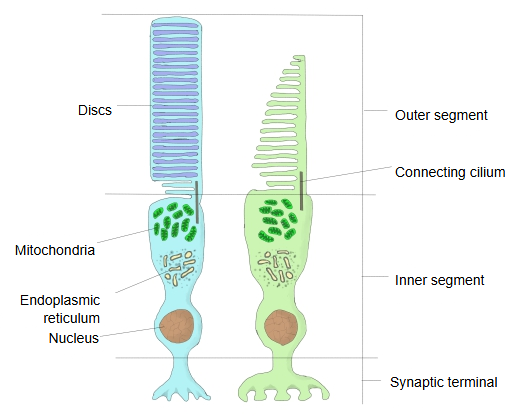
A rod is a long, thin cylinder. Its outer segment is a stack of flat membrane discs containing the visual pigment rhodopsin; its tip touches the pigment epithelium. A connecting cilium joins the outer segment to the inner segment, which holds the mitochondria and other organelles. The cell body with the nucleus lies in the outer nuclear layer, and the synaptic terminal (rod spherule) synapses with bipolar and horizontal cells.
Pathway
Rods and cones are the 1st neurons of the visual pathway; they turn light into electrical signals. The signal passes to bipolar cells (2nd neurons) and then to ganglion cells (3rd neurons), whose axons form the optic nerve. Many rods converge on one bipolar cell, which increases sensitivity in dim light but lowers sharpness.
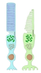 Photoreceptor cells (rod and cone)
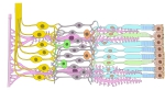 Structure of the retina
- 1st to 3rd neurons
Reference websites
[Wikipedia]
· Rod cell, Wikipedia.
· Photoreceptor cell, Wikipedia.
[Websites with illustrations]
· Rod cell (3-D drawing) (doctorc.net)

· Rod cell (illustration) (cdn.shopify.com)

· Retina diagram (Wikimedia Commons)

Related terms
- Retina
- Outer segment of photoreceptor
- Inner segment of photoreceptor
- Retinal pigment epithelium
- Optic part of retina
- Fovea centralis
- Macula lutea
References and sources
- Further reading: Rod cell, Wikipedia.
- Further reading: Photoreceptor cell, Wikipedia.
- Federative International Programme for Anatomical Terminology (FIPAT). Terminologia Anatomica, 2nd ed. (TA2), 2019.