Fovea centralis (Latin: fovea centralis)
Also called the central fovea or the fovea
· The fovea centralis is a depression at the centre of the macula lutea; its central part (the foveal avascular zone) has no retinal blood vessels.
· Visual acuity is highest at the fovea; vision with this part is called central vision.
· The fovea lies at about the posterior pole of the eyeball.
· The centre of the fovea is called the foveola; it is made only of cone cells (cone photoreceptors) and has no rods.
Note: the diameter differs a great deal between sources, probably because the anatomical and clinical definitions of the fovea are not the same.
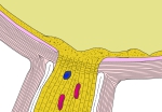 Optic disc and fovea (section)
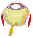 Right eyeball (horizontal section)
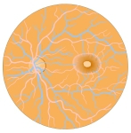 Left fundus
 Optic disc and macula
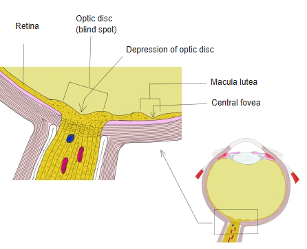
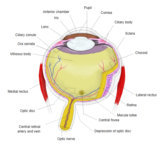
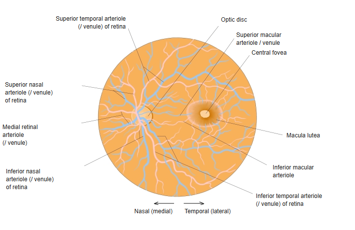
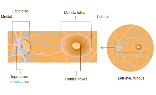
A description of how the fovea looks in the living eye:
In a fresh eye, and therefore also in the living eye seen with an ophthalmoscope, the macula lutea and the fovea look not yellow but reddish brown
to brown. The floor of the fovea is thin and transparent, so the layers beneath it show through faintly.
Reference websites
[Wikipedia]
· Fovea centralis, Wikipedia.
[Websites with illustrations]
· Macula and fovea of the eye (Cleveland Clinic)
· Eye anatomy with the macula and fovea (Wikimedia Commons)

Structure
The fovea is a depression in the inner surface of the retina, about 1.5 mm wide; its photoreceptor layer is made almost entirely of cones (only its centre, the foveola, is completely free of rods), and it is specialized for the highest visual acuity. Within it lies the foveal avascular zone, about 0.5 mm across, which has no retinal blood vessels; the fovea receives its oxygen from the vessels of the choroid beneath the retinal pigment epithelium. The inner layers of the retina are pushed aside to the rim of the pit, so light reaches the photoreceptors directly; the rim is the thickest part of the retina. The centre of the fovea, the foveola (about 0.35 mm across), contains only very densely packed, slender cones; toward the edge of the fovea rods appear and the density of cones falls. The fovea is surrounded by a ring called the parafovea and an outer ring called the perifovea, where acuity falls off. Although small, it is the only part of the retina that gives full visual acuity and the best colour vision, and about half of the fibres of the optic nerve carry information from it.
Related terms
References and sources
- Further reading: Fovea centralis, Wikipedia.
- Federative International Programme for Anatomical Terminology (FIPAT). Terminologia Anatomica, 2nd ed. (TA2), 2019.