Myoepithelial cell
Also called the basket cell (of glands)
· Their cytoplasm is rich in alpha-smooth muscle actin (αSMA), together with myosin, as in a smooth muscle cell; when it contracts, the flat, branched cells squeeze the acini and small ducts they wrap like a basket,
and this is thought to help the exocrine gland to release its secretion. The contraction is triggered by nerves or hormones; in the breast, oxytocin makes them contract and eject milk into the lactiferous ducts.
· Some myoepithelial cells form the basal layer of the epithelium and include the progenitor cells and stem cells of the gland epithelium.
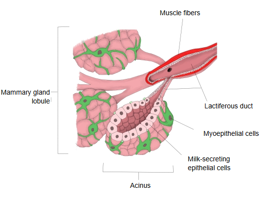
More about myoepithelial cells:
Myoepithelial cells lie between the glandular epithelium lining the acini and ducts of exocrine glands and the basement membrane. They are true epithelial cells (they contain keratin), but their α-smooth muscle actin lets them contract and help push the secretion out. They are found in the sweat, mammary, lacrimal and salivary glands and in the glands of the bronchi and oesophagus. Some of them form the basal layer of the epithelium and include its progenitor and stem cells; in wound healing they multiply to make up for lost cells. In pathology, the presence of myoepithelial cells in a proliferating tissue suggests a benign lesion, and their absence supports a malignant tumour; rare tumours such as adenomyoepithelioma, adenoid cystic carcinoma and myoepithelial carcinoma, however, contain tumorous myoepithelial cells as one of their components.
Most of the bipolar bare nuclei seen in cytology specimens of a fibroadenoma of the breast come from myoepithelial cells.
Markers
Myoepithelial cells are true epithelial cells that stain for keratin; they must not be confused with myofibroblasts, which are true mesenchymal cells that stain for vimentin. Myoepithelial cells usually express α-smooth muscle actin (αSMA), cytokeratins 5/6 and 14 and other high-molecular-weight cytokeratins, p63, CD10, calponin and caldesmon.
Reference websites
[Wikipedia]
· Myoepithelial cell, Wikipedia.
[Websites with illustrations]
· Myoepithelial cells in the breast (figure) (Springer Nature (Breast Cancer Research))

· Myoepithelial cells (figure) (Springer Nature)

Related terms
- Exocrine gland
- Basement membrane
- Sweat gland
- Lactiferous duct
- Lobes of mammary gland
- Acinar cells
- Mammary gland
References and sources
- Further reading: Myoepithelial cell, Wikipedia.
- Federative International Programme for Anatomical Terminology (FIPAT). Terminologia Anatomica, 2nd ed. (TA2), 2019.