Major salivary glands (Latin: glandulae salivariae majores)
The major salivary glands are the salivary glands that lie outside the oral mucosa: the parotid, submandibular and sublingual glands.
Glands that secrete saliva (most of it containing both mucus and serous fluid)
Major
salivary
glands
salivary
glands
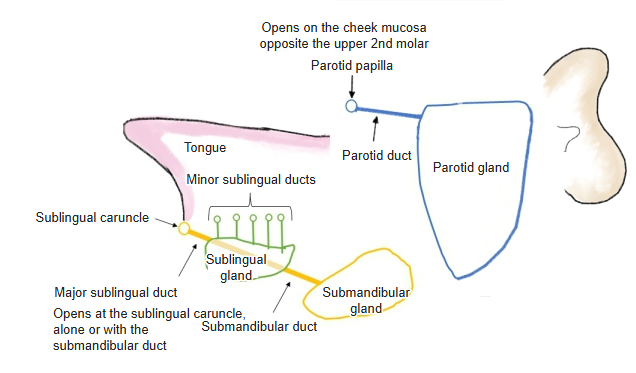
- Parotid gland : a flattened, irregular triangular gland in front of and below the external acoustic opening; the largest salivary gland; serous. The parotid duct opens at the parotid papilla. Share of unstimulated saliva: about 20%, rising to about 50% when stimulated.
- Submandibular gland : a flattened oval gland in the submandibular triangle; mixed. The submandibular duct opens on the sublingual caruncle. Its share of unstimulated saliva is the largest: about 60-67% (some sources give 75%).
- Sublingual gland : in the floor of the mouth, long from front to back and flattened from side to side; the smallest; mixed. The major sublingual duct opens on the sublingual caruncle, the minor sublingual ducts along the sublingual fold.
- Common feature : they lie at a distance from the oral mucosa and release saliva through ducts.
· They differ from the minor salivary glands, which lie in the mucosa itself and open directly or by very short ducts.
· Together they produce most of the saliva; the submandibular glands give the largest share at rest, and the parotid glands increase their output most when stimulated by eating. Sources differ on the exact shares.
· Their secretory (parasympathetic) supply comes from the facial nerve (submandibular and sublingual glands) and the glossopharyngeal nerve (parotid gland).
Salivary
glands
glands
- Major salivary glands : lie at a distance from the oral mucosa and release saliva through ducts.
- Parotid gland : an irregular, flattened triangular gland in front of and below the external acoustic meatus; the largest; serous. The parotid duct opens into the vestibule of the mouth.
- Submandibular gland : a flattened oval gland in the submandibular triangle; mixed. The submandibular duct opens on the sublingual caruncle.
- Sublingual gland : in the floor of the mouth, long from front to back and flattened from side to side; the smallest; mixed.
- Minor salivary glands : lie in the mucosa and release saliva into the mouth directly or through short ducts.
- Labial glands : in the mucosa of the lips; mixed. Few at the corners of the mouth; some lie in the muscle layer.
- Buccal glands : in the mucosa of the cheeks; mixed. Some also lie outside the buccinator.
- Molar glands : at the back of the cheek, between the masseter and the buccinator; mixed. They open opposite the last upper molar teeth.
- Palatine glands : in the back of the hard palate and throughout the soft palate (sources differ on where they are densest); mucous.
- Lingual glands : three groups: the anterior lingual glands (mixed) under the tip of the tongue, opening beside the lingual frenulum; the posterior lingual glands (mucous) in the root and along the back of the margins; the glands of von Ebner (serous), opening into the trenches of the vallate and foliate papillae.
Reference websites
[Wikipedia]
· Salivary gland, Wikipedia.
[Websites with illustrations]
· The salivary glands (TeachMeAnatomy)
· The oral cavity (TeachMeAnatomy)
Related terms
- Salivary glands
- Parotid gland
- Submandibular gland
- Sublingual gland
- Minor salivary glands
- Parotid papilla
- Sublingual caruncle
References and sources
- Further reading: Salivary gland, Wikipedia.
- Federative International Programme for Anatomical Terminology (FIPAT). Terminologia Anatomica, 2nd ed. (TA2), 2019.