Retinal ganglion cell
Also called Ganglion cell of retina or RGC
The main facts:
· It carries the visual information of the retina to the thalamus, the hypothalamus and the midbrain (it has a long axon that reaches the brain).
· The human retina has roughly 0.7–1.5 million ganglion cells (sources differ).
(There are about 100 million photoreceptor cells, so on average one ganglion cell receives input from about 100 cone
and rod cells.)
(In the fovea the convergence is smallest: a ganglion cell receives from only a few photoreceptors.)
(At the extreme periphery of the retina, one ganglion cell receives information from thousands of photoreceptor cells.)
· It produces action potentials spontaneously, even at rest.
· From the pattern of their projections and the properties of their receptive fields, at least five main types of ganglion cell are thought to exist.
The path of the visual signal in the retina
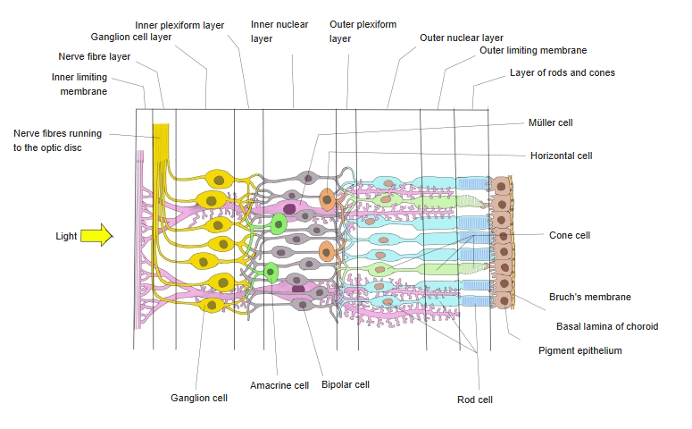
Light reaching the retina is first sensed by the photoreceptor cells (rods and cones), the first neurons, which lie in the outermost layer of the retina.
of the
optic part
of the
retina
- Photoreceptor cells
- Rod cell : rod-shaped outer segment; most photoreceptors are rods. They serve light reception in dim light.
- Cone cell : cone-shaped outer segment; concentrated in the centre of the retina. They work in bright light and give sharp and colour vision.
- Horizontal cell : its processes spread sideways along the retina and synapse with the photoreceptor cells.
- Bipolar cell : has two processes, one on each side of the cell body; passes the signal from the photoreceptors to the ganglion cells.
- Amacrine cell : a local interneuron without a long axon; links bipolar cells and ganglion cells.
- Ganglion cell : has a long axon that runs to the brain; the axons form the optic nerve, optic chiasm and optic tract.
- Müller cell : not a neuron but a kind of glial cell; it spans the retina as a supporting cell and does not carry the visual signal.
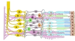 Structure of the retina
- 1st to 3rd neurons
Reference websites
[Wikipedia]
· Retinal ganglion cell, Wikipedia.
· Retina, Wikipedia.
[Websites with illustrations]
· Plan of the retinal neurons (Gray's Anatomy) (Wikimedia Commons)

· Retina (Kenhub)
Structure and function
The cell bodies lie in the ganglion cell layer, near the inner surface of the retina facing the vitreous body. Their axons run in the nerve fibre layer to the optic disc, where they leave the eye as the optic nerve. They receive signals from the rods and cones through the bipolar cells, and amacrine and horizontal cells modify the signal on the way. Unlike the rods, cones and bipolar cells they fire action potentials, at a base rate even at rest: excitation raises the rate and inhibition lowers it. The axons end mainly in the lateral geniculate body of the thalamus, with smaller branches to the hypothalamus (body clock) and the midbrain (pupil reflex and eye movements). The numbers of photoreceptors per ganglion cell vary greatly between people and with the part of the retina.
Related terms
References and sources
- Further reading: Retinal ganglion cell, Wikipedia.
- Federative International Programme for Anatomical Terminology (FIPAT). Terminologia Anatomica, 2nd ed. (TA2), 2019.