Ganglion (Latin: ganglion)
Also called Plural: ganglia
- Overview
- Sensory ganglia (Spinal (dorsal root) ganglion, Main sensory ganglia of the cranial nerves, Structures called ganglia that are not true ganglia)
- Reference websites
- (Wikipedia, websites with illustrations)
- Related terms
Overview
· The swelling is due to the densely packed cell bodies; where synapses are present, they also add to it.
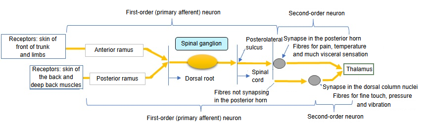
- Sensory ganglia : first-order sensory neurons, mostly pseudounipolar (bipolar in the cochlear and vestibular ganglia); no synapses
- Spinal (dorsal root) ganglia : on the dorsal roots of the spinal cord; 31 on each side, 62 in all
- Ganglia of the cranial nerves : e.g. the trigeminal ganglion, with the same basic structure as the spinal ganglia, and the cochlear (spiral) ganglion, whose neurons are bipolar
- Autonomic ganglia : multipolar neurons; synapses from preganglionic fibres onto the neurons of the postganglionic fibres
- Sympathetic ganglia
- Paravertebral ganglia : on both sides of the vertebral column = ganglia of the sympathetic trunk
- Prevertebral ganglia : around the abdominal aorta and its branches = preaortic ganglia
- Parasympathetic ganglia : small ganglia near or within the target organs
- Sympathetic ganglia
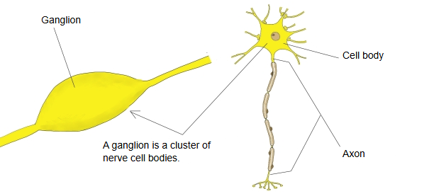
Sensory ganglia
Spinal (dorsal root) ganglion
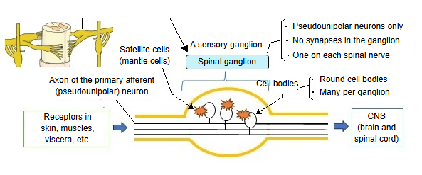
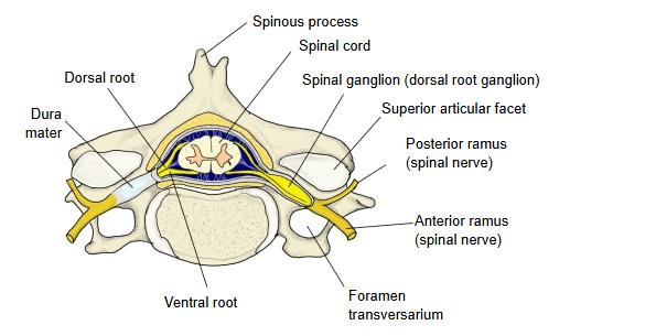
The ganglion on the dorsal root of each spinal nerve (spinal cord). Of the two roots, only the dorsal root carries a ganglion, so the term means the same as spinal ganglion.
There is one for each spinal nerve: 31 on each side, 62 in all (there are 31 pairs of spinal nerves).
· Each ganglion is a small swelling on the dorsal root, lying in or near the intervertebral foramen.
· The cell bodies of the sensory (pseudounipolar) neurons are round and vary in size; each is surrounded by satellite cells.
· Each ganglion contains a very large number of cell bodies.
Published sizes and cell counts differ widely between sources.
The size of the dorsal root ganglia differs by region. The table gives the general pattern in the adult.
| Region | Size | Notes | |
|---|---|---|---|
| 1 | Cervical (C5-C8) | Larger | Collect much sensory input from the upper limb |
| 2 | Thoracic (T1-T12) | Smaller | Mainly the skin of the chest and trunk wall |
| 3 | Lumbar (L1-L5) | Larger, especially at the lower lumbar levels | Collect much sensory input from the lower limb |
| 4 | Sacral (S1-S5) | Larger at the upper sacral levels, smaller below | The pelvis and the distal parts of the lower limb |
| Name | Notes | |
|---|---|---|
| 1 | Trigeminal ganglion | Nerve: trigeminal nerve (CN V) Sensation: touch, pain and temperature of the face Neurons: mainly pseudounipolar |
| 2 | Superior cervical ganglion | Sometimes mentioned with the head ganglia, but it is not a sensory ganglion: it is the uppermost ganglion of the sympathetic trunk, whose fibres reach the head |
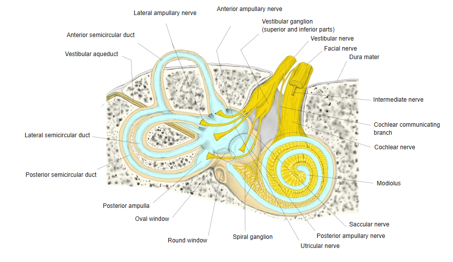
| 3 | Vestibular ganglion | Nerve: vestibular nerve (CN VIII) Sensation: balance Neurons: bipolar |
| 4 | Cochlear (spiral) ganglion | Nerve: cochlear nerve (CN VIII) Sensation: hearing Neurons: bipolar |
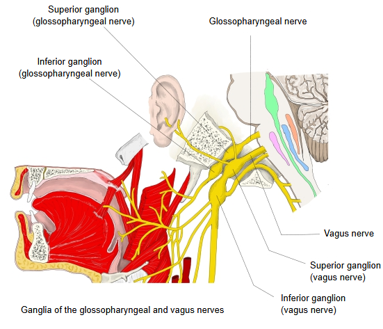
| 5 | Ganglia of the glossopharyngeal nerve | Superior ganglion / inferior ganglion Sensation: taste from the posterior third of the tongue; sensation of the pharynx |
| 6 | Ganglia of the vagus nerve | Superior ganglion / inferior ganglion Sensation: pharynx, larynx and viscera |
· Sensory ganglia hold the cell bodies of the sensory neurons that carry information from receptors in the periphery to the central nervous system.
· In form, sensory neurons are bipolar or pseudounipolar.
Main sensory ganglia of the cranial nerves
The table lists the sensory ganglia of the cranial nerves as fully as possible, but it may not be complete.
| Name | Other name | Afferent nerve | Location | |
|---|---|---|---|---|
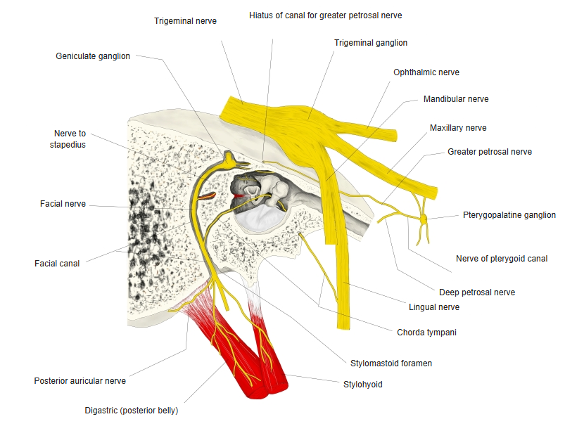
| 1 | Trigeminal ganglion | Semilunar ganglion | Ophthalmic, maxillary and mandibular nerves | On the trigeminal impression near the apex of the petrous part of the temporal bone |
| 2 | Geniculate ganglion | Facial nerve | At the first bend of the facial nerve (geniculum of facial nerve) | |
| 3 | Vestibular ganglion | Scarpa's ganglion | Vestibular nerve (vestibulocochlear nerve) | In the internal acoustic meatus, near its fundus |
| 4 | Spiral ganglion | Cochlear ganglion | Cochlear nerve (vestibulocochlear nerve) | In the modiolus of the cochlea |
| 5 | Superior ganglion of glossopharyngeal nerve | Superior glossopharyngeal ganglion | Glossopharyngeal nerve | Upper part of the jugular foramen |
| 6 | Inferior ganglion of glossopharyngeal nerve | Inferior glossopharyngeal ganglion | Glossopharyngeal nerve | Lower part of the jugular foramen |
| 7 | Superior ganglion of vagus nerve | Jugular ganglion | Vagus nerve | In the jugular foramen |
| 8 | Inferior ganglion of vagus nerve | Nodose ganglion | Vagus nerve | Just below the jugular foramen |
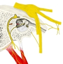 Trigeminal ganglion / geniculate ganglion
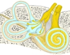 Vestibular ganglion / spiral ganglion
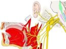 Ganglia of the glossopharyngeal and vagus nerves
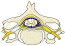 Spinal (dorsal root) ganglion
Structures called ganglia that are not true ganglia
· Basal ganglia: the term ganglion strictly belongs to the peripheral nervous system. The basal ganglia are groups of nuclei deep in the cerebrum, in the central nervous system, connected with the cerebral cortex, thalamus and brainstem; they take part in the control of movement and in cognition, emotion and learning. The anatomical terminology therefore recommends the name basal nuclei, although basal ganglia is still widely used.
· Pseudoganglion: a local thickening of a nerve that looks like a ganglion but contains only nerve fibres, with few or no nerve cell bodies. Examples are described on the nerve to teres minor and on the radial nerve.
Reference websites
[Wikipedia]
· Ganglion, Wikipedia.
· Dorsal root ganglion, Wikipedia.
· Cranial nerve ganglia, Wikipedia.
· Autonomic ganglion, Wikipedia.
[Websites with illustrations]
· Dorsal root ganglion (Kenhub)
· Formation of a spinal nerve from the roots of the spinal cord (Gray's Anatomy) (Wikimedia Commons)

Related terms
References and sources
- Further reading: Ganglion, Wikipedia.
- Further reading: Cranial nerve ganglia, Wikipedia.
- Further reading: Autonomic ganglion, Wikipedia.
- Federative International Programme for Anatomical Terminology (FIPAT). Terminologia Anatomica, 2nd ed. (TA2), 2019.