Cholangiocyte
Also called the biliary epithelial cell or the bile duct epithelial cell
· The epithelium of cholangiocytes that lines the bile ducts is always simple (a single layer).
· The shape of the cells changes along the ducts: simple squamous ⇒ simple cuboidal ⇒ simple columnar, becoming taller as the ducts get wider.
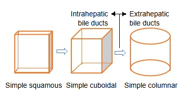
The epithelium along the bile pathway, part by part:
| Part | Type of epithelium | Cell shape | Notes |
|---|---|---|---|
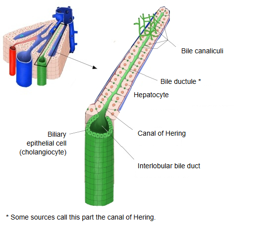
| Bile canaliculus | No epithelium of its own; a groove between hepatocytes | Where the bile begins, between the hepatocytes | |
| Canal of Hering | Simple squamous to cuboidal epithelium | Low cuboidal to flat | The junction between the bile ducts and the hepatocytes; lined partly by hepatocytes |
| Interlobular bile duct | Simple cuboidal epithelium | Small cuboidal cells | In the portal tracts |
| Segmental bile ducts to the right and left hepatic ducts | Simple columnar epithelium | Cells become taller | The bile ducts become wider and the cells more active in secretion |
| Extrahepatic bile ducts | Simple columnar epithelium | Tall columnar | Mucous glands in the wall and some mucus-secreting cells; each cell has a single primary cilium |
Functions
Final adjustment of the quality and quantity of bile
· Cholangiocytes add bicarbonate (HCO₃⁻), water and electrolytes to the primary bile made by the hepatocytes, keeping the bile alkaline.
Main molecules: CFTR (a Cl⁻ channel), AE2 (a Cl⁻/HCO₃⁻ exchanger) and aquaporin water channels.
· When needed they reabsorb glucose, amino acids, bile acids and other substances, fine-tuning the composition of the bile.
· In this way they help fat digestion in the intestine and form a protective "bicarbonate umbrella" that shields the epithelial cells from damage by bile acids.
Control of bile flow (choleresis)
· They respond to hormones, autonomic nerves and bile-acid signals, increasing or decreasing their secretion through the cAMP and Ca²⁺ pathways.
Example: secretin → cAMP rises → CFTR is activated → secretion of Cl⁻ and HCO₃⁻ increases.
Stimulation by the vagus nerve and other parasympathetic fibres (acetylcholine) acts mainly through the Ca²⁺ pathway.
Protective barrier and immune regulation
· They secrete mucins, IgA and antimicrobial peptides, which protect the duct lining from bacteria and toxins flowing back from the intestine.
· They produce cytokines and chemokines (such as IL-6 and IL-8) that recruit immune cells in the liver.
· They express HLA class I and II molecules and, under some conditions, can act as antigen-presenting cells.
Support for metabolism and detoxification
· They express low levels of CYP enzymes, glucuronosyltransferases and other enzymes, helping to detoxify drugs and bile acids.
· Organic anion transporters (OATP/OAT, MRP and others) carry bile components and drug metabolites.
Regeneration and differentiation
· Small cholangiocytes multiply as stem or progenitor cells after liver injury and can differentiate into hepatocytes and large cholangiocytes (according to some sources).
· They secrete VEGF, TGF-β and other factors, helping to form the microenvironment around the bile ducts and portal vein branches (the biliary niche).
A hub of signalling
· They secrete ATP, nitric oxide (NO) and exosomes, which act on nearby hepatic stellate cells and vascular endothelium (paracrine action).
· With their primary cilium they sense the mechanical stimulus of bile flow and turn it into a Ca²⁺ signal, acting as "mechanosensors".
Reference websites
[Wikipedia]
· Cholangiocyte, Wikipedia.
· Bile duct, Wikipedia.
[Websites with illustrations]
· Cholangiocyte functions (figure) (Nature Reviews Gastroenterology & Hepatology)

· The biliary tree and cholangiocytes (figure) (Nature Reviews Gastroenterology & Hepatology)

Related terms
- Bile duct
- Interlobular bile duct
- Canal of Hering
- Bile ductule
- Hepatocyte
- Common bile duct
- Epithelial cell
References and sources
- Further reading: Cholangiocyte, Wikipedia.
- Further reading: Bile duct, Wikipedia.
- Federative International Programme for Anatomical Terminology (FIPAT). Terminologia Anatomica, 2nd ed. (TA2), 2019.