Bile canaliculus (Latin: canaliculus biliferus)
Also called the bile capillary
Sources differ a good deal in the names of the channels that carry the bile made by the hepatocytes to the interlobular bile ducts outside the hepatic lobule, and it is not clear which naming is correct or most common. The description below is one example.
· The bile canaliculi form a branching network that runs lengthwise and crosswise inside the hepatic cell plates of the hepatic lobule.
· A bile canaliculus is only about 0.5–1.5 µm across.
· The bile canaliculus is also called the bile capillary.
The name "bile capillary" is sometimes used, but it is not a blood vessel; the standard anatomical term is bile canaliculus (Latin: canaliculus biliferus).
· Some textbooks use the name "bile capillary", while others use only "bile canaliculus".
The bile pathway inside the liver
Sources differ: some do not distinguish the bile canaliculus, canal of Hering and bile ductule, some do not use the names bile ductule and canal of Hering at all, and some reverse their order. The diameters are only rough guides.
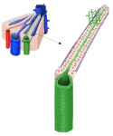 From bile canaliculi to bile duct
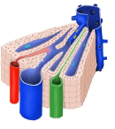 Structure of a hepatic lobule
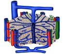 Hepatic lobule (cut-away)
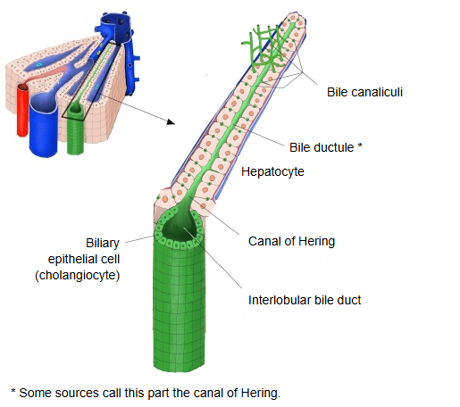
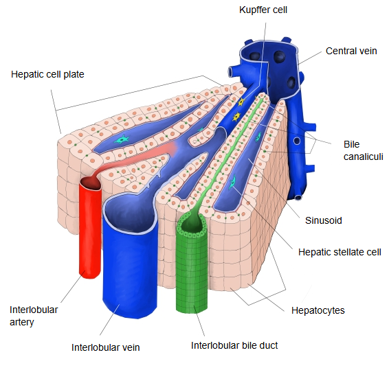
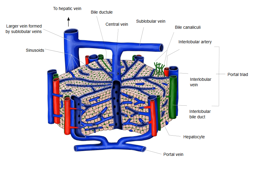
A bile canaliculus is a gap between hepatocytes with no special wall. It begins near the central vein, runs between the rows of liver cells to the edge of the hepatic lobule, and continues into a short connecting segment lined by simple cuboidal or flat cells, which is much wider than the canaliculus. Two or three of these segments join, leave the lobule and become an interlobular bile duct. Lymphatic vessels are also found in the interlobular connective tissue and Glisson's sheath. The bile canaliculus corresponds to the lumen of an ordinary gland, but unlike other glands it is bounded by only two hepatocytes.
Reference websites
[Wikipedia]
· Bile canaliculus, Wikipedia.
· Canals of Hering, Wikipedia.
[Websites with illustrations]
· Bile canaliculi and the hepatic lobule (figure) (DoctorLib)

Related terms
- Bile ductule
- Canal of Hering
- Interlobular bile duct
- Hepatocyte
- Hepatic cell plate
- Hepatic lobule
- Liver
References and sources
- Further reading: Bile canaliculus, Wikipedia.
- Further reading: Canals of Hering, Wikipedia.
- Federative International Programme for Anatomical Terminology (FIPAT). Terminologia Anatomica, 2nd ed. (TA2), 2019.