Pelvic floor muscles
Also called: muscles of the pelvic floor
The following seven muscles are listed:
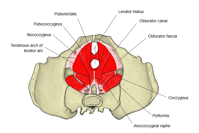
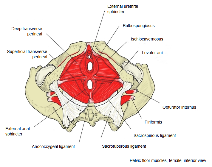
| 1 | Levator ani | one of the anal muscles; it has three parts: the puborectalis, the pubococcygeus and the iliococcygeus |
| 2 | Superficial transverse perineal | one of the urogenital muscles |
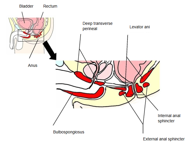
| 3 | Deep transverse perineal | one of the urogenital muscles |
| 4 | External anal sphincter | one of the anal muscles |
| 5 | External urethral sphincter | one of the urogenital muscles; also called the sphincter urethrae |
| 6 | Bulbospongiosus | one of the urogenital muscles |
| 7 | Ischiocavernosus | one of the urogenital muscles |
This list follows one anatomy atlas. "Pelvic floor muscles" is not an official anatomical term, and some textbooks do not use it. Some textbooks use the name muscles of the perineum instead; almost all of these (all except the coccygeus) are pelvic floor muscles. In the narrow sense the term means only the pelvic diaphragm (levator ani and coccygeus).
- Urogenital group : muscles of the external genitalia
- Bulbospongiosus : pelvic floor muscle
- Ischiocavernosus : pelvic floor muscle
- Superficial transverse perineal : pelvic floor muscle
- Deep transverse perineal : pelvic floor muscle
- External urethral sphincter : pelvic floor muscle (sphincter urethrae)
- Anal group
- Levator ani : pelvic floor muscle: puborectalis, pubococcygeus, iliococcygeus
- External anal sphincter : pelvic floor muscle
- Coccygeus : not counted here
Other sources name only the following four groups as the pelvic floor muscles:
· the deep transverse perineal · the sphincter urethrae · the anal muscles · the coccygeus
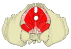 Female pelvic floor (superior view)
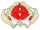 Female pelvic floor (inferior view)
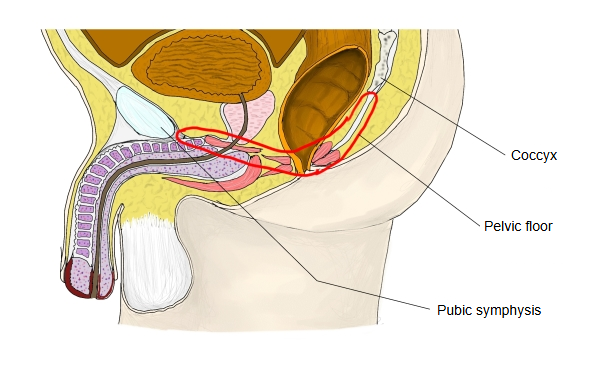
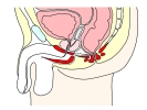 Pelvic floor (median section)
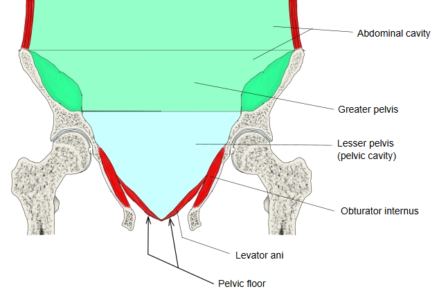
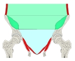 Pelvis (coronal section)
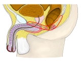 Pelvic floor (median section)
Functions
The pelvic floor muscles have the following functions.
(1) Support of the organs of the abdominal cavity and the pelvic cavity
Stretched like a hammock across the floor of the pelvis, they hold the intestines, the urinary bladder and the other organs of the abdominal and pelvic cavities in their proper places against the pressure inside the abdomen.
(2) Control of excretion of urine and faeces
Through their sphincter muscles they tighten or relax the openings of the urethra and the rectum and anal canal, and so control the passing of urine and faeces.
To do these opposite jobs (closing the pelvic cavity while keeping several openings), the pelvic floor is lined by overlapping layers of connective tissue. This complex structure makes it easily damaged, especially in women. Repeated rises in pressure inside the abdomen and other strains, above all late in pregnancy, weaken its connective tissue and its muscles, and stretching or injury during childbirth can lead in the end to failure of the pelvic floor. The results include descent of the pelvic organs (for example of the uterus), in severe cases prolapse of the uterus with the vagina turned outwards, and loss of urine or faeces on coughing or straining (stress incontinence). Mild cases often improve with regular pelvic floor exercises; severe cases often need surgical repair of the pelvic floor, in which the two limbs of the levator ani (the puborectalis) are exposed and brought together.
Reference websites
[Wikipedia]
· Pelvic floor, Wikipedia.
[Websites with illustrations]
· Pelvic diaphragm, female (illustration) (Netter Images)

· The pelvic floor: structure, function, muscles (TeachMeAnatomy)
Related terms
References and sources
- Further reading: Pelvic floor, Wikipedia.
- Federative International Programme for Anatomical Terminology (FIPAT). Terminologia Anatomica, 2nd ed. (TA2), 2019.