Thick ascending limb (Latin: pars recta tubuli distalis)
Also called the distal straight tubule (TAL)
Overview
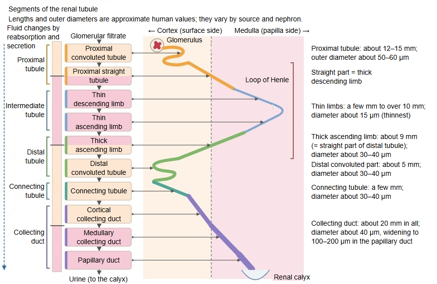
· The thick ascending limb is the part of the ascending limb of the loop of Henle that has a tall epithelium. It runs straight up from the outer zone of the renal medulla through a medullary ray of the renal cortex, touches the vascular pole of its own renal corpuscle and then continues as the distal convoluted tubule. It is also called the distal straight tubule, the straight part of the distal tubule.
· It is divided into a medullary part, in the renal medulla, and a cortical part, in the renal cortex. In cortical nephrons it follows the thin descending limb; in juxtamedullary nephrons it follows the thin ascending limb.
· Its wall is a simple cuboidal epithelium without a brush border. The cells are rich in mitochondria and have deep folds of the basal membrane (basal infoldings); the nuclei bulge towards the lumen.
· It is almost impermeable to water but actively reabsorbs sodium, potassium and chloride ions through the Na–K–2Cl cotransporter (NKCC2); about 25% of the filtered sodium is reabsorbed here. Calcium and magnesium are also reabsorbed, through the spaces between the cells. Because salt is returned without water, the fluid in the tubule becomes dilute (this segment is also called the diluting segment) and the surrounding medulla becomes hyperosmotic.
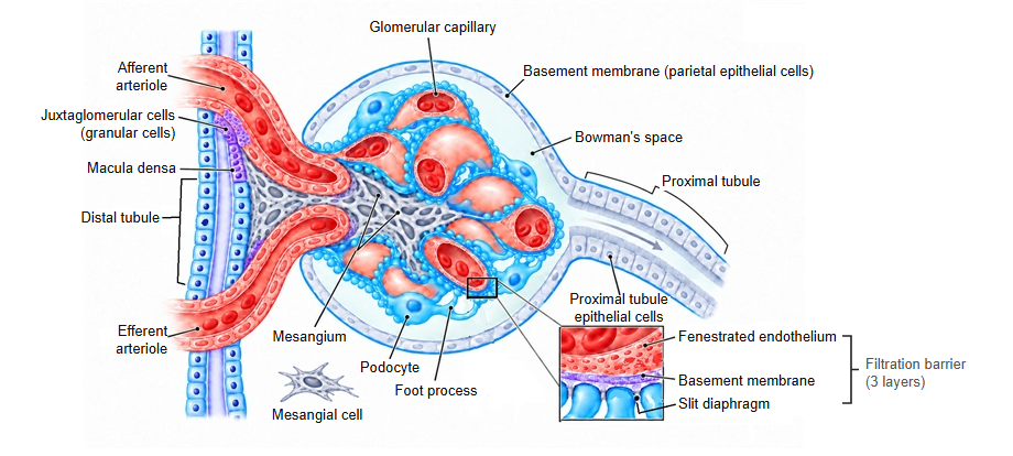
· Where the thick ascending limb touches the vascular pole, its epithelium is tall and closely packed and forms the macula densa. As part of the juxtaglomerular apparatus, the macula densa senses the sodium chloride concentration of the tubular fluid and helps to regulate the glomerular filtration rate.
· Loop diuretics such as furosemide block NKCC2 and increase urine output. Uromodulin (Tamm–Horsfall protein), the most abundant protein in normal urine, is made by the epithelium of this segment.
Reference websites
[Wikipedia]
· Loop of Henle, Wikipedia.
[Websites with illustrations]
· Tubules and collecting system (16) (Digital Histology)
· Tubules and collecting system (15) (Digital Histology)
· Tubules and collecting system (8) (Digital Histology)
· Molar transport along the nephron (Wikimedia Commons)

Related terms
- Loop of Henle
- Distal tubule
- Straight tubule
- Macula densa
- Juxtaglomerular apparatus
- Medullary rays
- Vascular pole
References and sources
- Further reading: Loop of Henle, Wikipedia.
- Federative International Programme for Anatomical Terminology (FIPAT). Terminologia Anatomica, 2nd ed. (TA2), 2019.