Area cribrosa (Latin: area cribrosa)

Overview
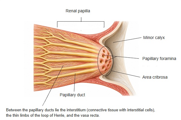
The area cribrosa is the part at the tip of the renal papilla where many papillary foramina open, so that it looks like a sieve.
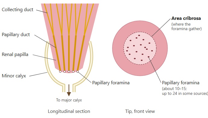
About 10–15 foraminaA sieve is a tool with many small holes
Primary urine is discharged from the papillary foramina into the minor calyx and becomes urine. In other words, the area cribrosa is the boundary between primary urine and urine.
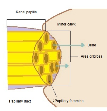
Left, a small schematic of a nephron and collecting duct with a box at the tip of the papilla; centre, that tip enlarged: papillary ducts end as papillary foramina in the area cribrosa, which projects into the minor calyx, and urine flows out into the calyx; right, a woven sieve for comparison.
The tip of the renal papilla, and a sieve (click to enlarge)

· Seen from the surface, a renal papilla has about 10 to 15 papillary foramina (larger papillae up to about 24). The openings can be seen with the naked eye and give the surface a sieve-like look, which is why this part is called the area cribrosa. The foramina are the outlets of the papillary ducts, which are formed by the union of collecting ducts.

· The papilla, with the area cribrosa at its tip, projects into the minor calyx, and the rim of the minor calyx is attached around the renal papilla.

· In a compound papilla, formed by the fusion of two or three papillae, the area cribrosa is larger and the papillary foramina are more numerous.

· Cribrosa is Latin for "sieve-like"; it is the same word as in lamina cribrosa, the cribriform plate of the ethmoid bone.
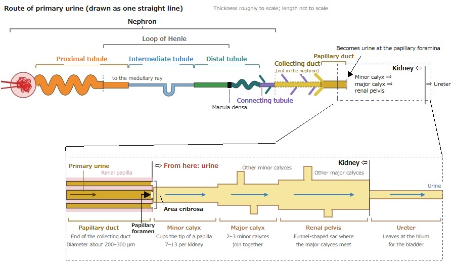
Upper part: the route of primary urine drawn as one straight line, from the renal corpuscle through the proximal, intermediate and distal tubules, the connecting tubule, collecting duct and papillary duct to the calyces, renal pelvis and ureter. Lower part, enlarged: primary urine in the papillary duct passes the papillary foramen at the area cribrosa and from there is urine, flowing through the minor calyx, major calyx and renal pelvis to the ureter.
Route of primary urine (schematic) (click to enlarge)
Schematic. Left, a longitudinal section: collecting ducts join into papillary ducts that open as papillary foramina at the tip of the renal papilla, which projects into the cup of a minor calyx leading on to a major calyx. Right, the tip seen from the front: the dotted area cribrosa in the middle, where about 10 to 15 papillary foramina open.
Renal papilla, papillary foramina and area cribrosa (schematic) (click to enlarge)
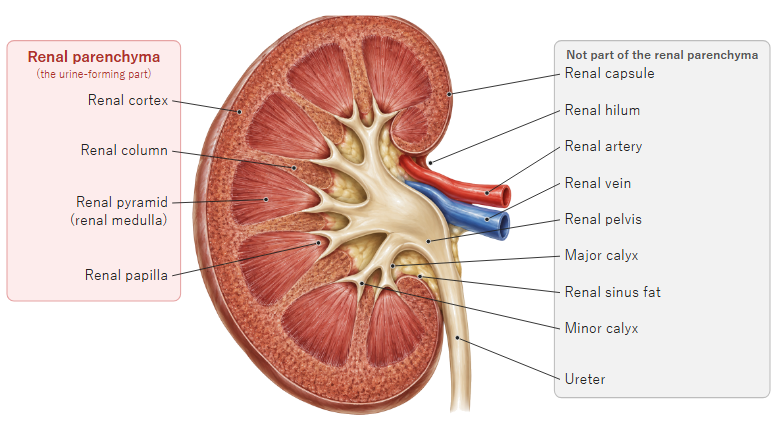
A coronal section of a kidney. Labels on the left mark the renal cortex, renal column, renal pyramid, renal medulla and renal papilla; labels on the right mark the renal capsule, hilum, renal artery and vein, renal pelvis, major and minor calyces and ureter.
Renal parenchyma (coronal section of the kidney) (click to enlarge)
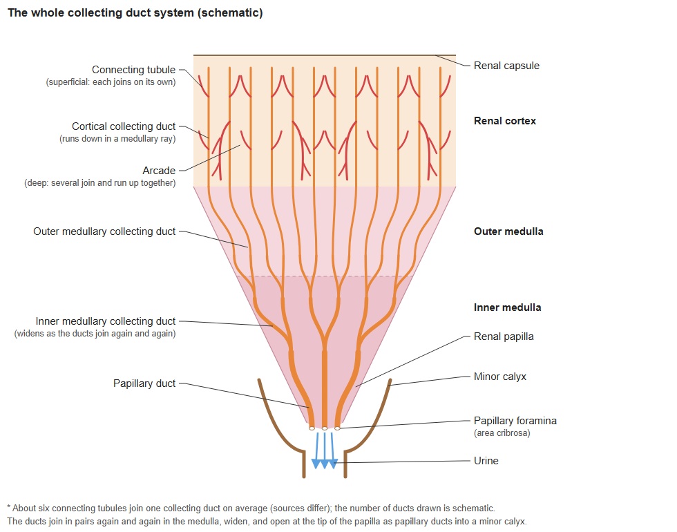
  • Small picture of the whole collecting duct from the cortex to the renal papilla; opens the labelled figure.The whole collecting duct
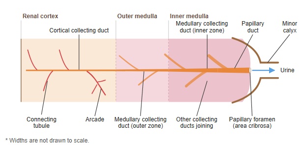
  • Small picture of the collecting duct drawn as one line from the cortex to the minor calyx; opens the labelled figure.Flow through the collecting duct
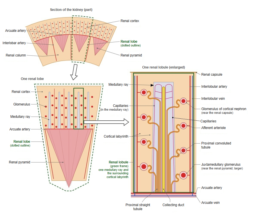
  • Small picture of a renal lobe and renal lobule; opens the labelled figure.Renal lobe and renal lobule
  • Small picture of a renal papilla projecting into a minor calyx; opens the labelled figure.Renal papilla

Reference websites

[Wikipedia]

· Renal papilla, Wikipedia.

· Renal calyx, Wikipedia.

[Websites with illustrations]

· Kidney histology (Kenhub)

· Section of the kidney (Gray's Anatomy, fig. 1127) (Wikimedia Commons)

· Kidney structure (labelled diagram) (Wikimedia Commons)

References and sources

  1. Further reading: Renal papilla, Wikipedia.
  2. Further reading: Renal calyx, Wikipedia.
  3. Federative International Programme for Anatomical Terminology (FIPAT). Terminologia Anatomica, 2nd ed. (TA2), 2019.