Papillary foramina (Latin: foramina papillaria)
Also called the openings of the papillary ducts
Overview
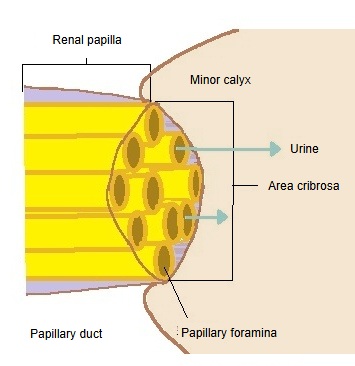
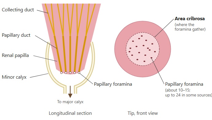
· The papillary foramina are the small openings on the tip of each renal papilla, where the papillary ducts, the last part of the collecting duct system, end. They are also called the openings of the papillary ducts.
· A small simple papilla has on average about 10 to 15 foramina, and a fairly large one up to 24, occasionally more. Compound papillae, formed when several papillae fuse, have even more.
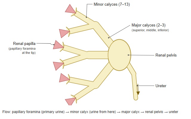
· Around the foramina, the columnar epithelium of the collecting duct system changes to the transitional epithelium (urothelium) that lines the inner surface of the minor calyx. The urine leaving the foramina passes in turn to a major calyx, the renal pelvis and the ureter.
· On an ordinary simple papilla the foramina are narrow slits that tend to close when pressure in the calyx rises. Compound papillae, which are common at the upper and lower poles of the kidney, may have round, open foramina; when there is vesicoureteral reflux, urine can then flow back into the renal parenchyma (intrarenal reflux). This is thought to be why the scars of reflux nephropathy tend to form at the poles.
 Schematic of the renal calyces
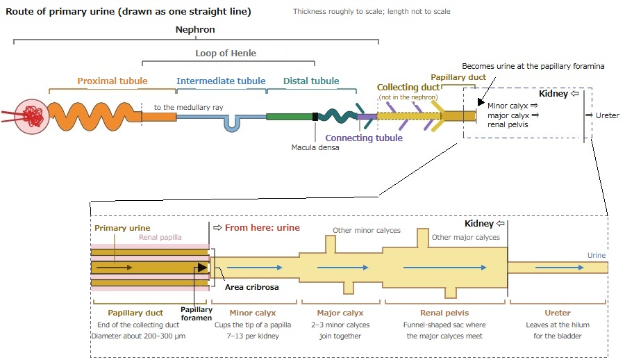
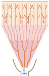 The whole collecting duct
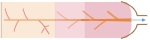 Flow through the collecting duct
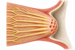 Renal papilla
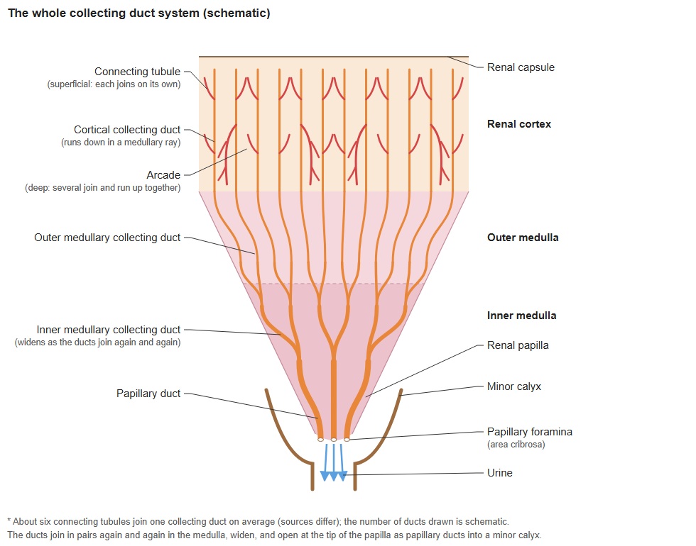
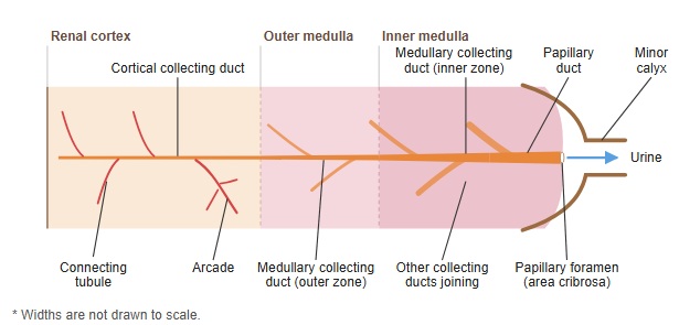
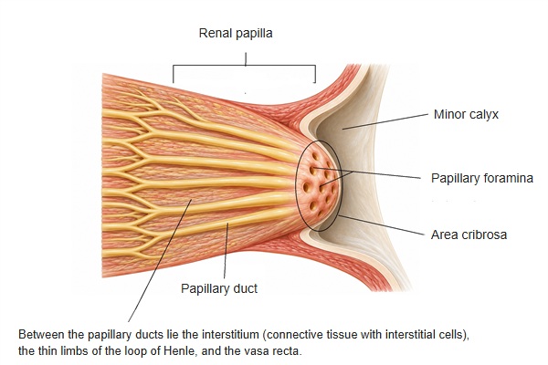
- Renal cortex
- Renal corpuscle : glomerulus and Bowman's capsule.
- Proximal tubule : takes back water and nutrients from the primary urine.
- Distal tubule : adjusts electrolytes and water.
- Cortical collecting duct : collects primary urine from several connecting tubules.
- Renal medulla
- Renal pyramid : the cone-shaped tissue that forms the medulla; it contains:
- Loop of Henle : part of the countercurrent system that lets the kidney concentrate urine.
- Medullary collecting duct : carries primary urine to the renal papilla.
- Papillary duct : opens at the tip of the papilla; its contents leave into a minor calyx as urine.
- Renal pyramid : the cone-shaped tissue that forms the medulla; it contains:
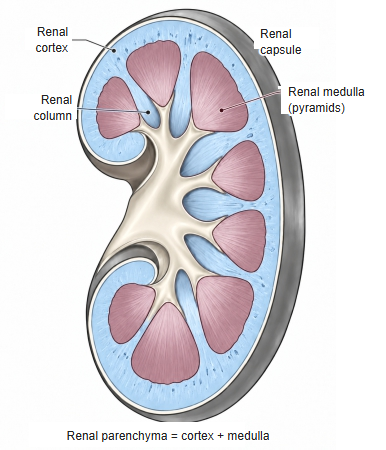
renal medulla (click to enlarge)
Reference websites
[Wikipedia]
· Renal papilla, Wikipedia.
· Collecting duct system, Wikipedia.
[Websites with illustrations]
· Kidney histology (Kenhub)
· Gray's Anatomy, fig. 1128 (Wikimedia Commons)

· The kidneys (TeachMeAnatomy)
Related terms
References and sources
- Further reading: Renal papilla, Wikipedia.
- Further reading: Collecting duct system, Wikipedia.
- Federative International Programme for Anatomical Terminology (FIPAT). Terminologia Anatomica, 2nd ed. (TA2), 2019.