Papillary duct (Latin: ductus papillaris)
Also called the duct of Bellini
Overview
- Cortical collecting duct : runs down in the medullary rays of the renal cortex.
- Medullary collecting duct : runs down through the renal medulla (outer and inner zones), joining with others and becoming wider.
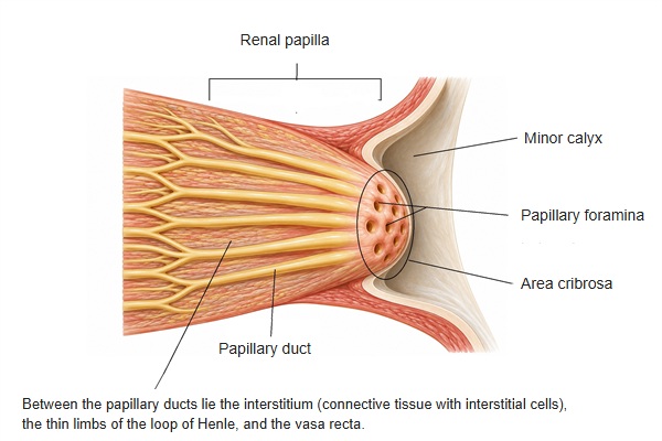
- Papillary duct : discharges primary urine from the papillary foramina at the tip of the renal papilla into the minor calyx, where it becomes urine.
Note: the papillary duct is sometimes counted as part of the medullary collecting duct, as the wide final part of the inner medullary collecting duct.
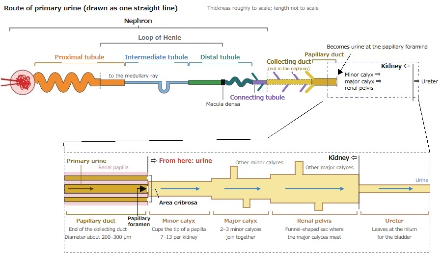
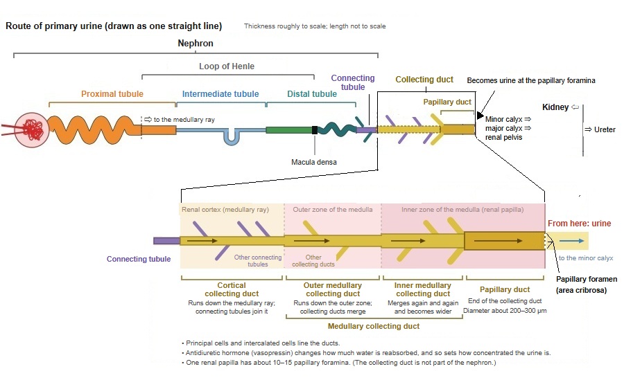
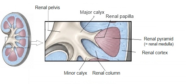
· The papillary ducts open at the apex of the renal pyramid (the renal papilla) as many papillary foramina; the part where the foramina gather is called the area cribrosa.
· The primary urine carried by the papillary ducts flows out into a minor calyx, where it becomes urine.
· The wall is tall columnar epithelium with light principal cells and a few dark intercalated cells. Near the minor calyx the lining changes to urothelium (transitional epithelium).
· Water reabsorption and electrolyte handling continue here under hormones such as vasopressin and aldosterone.
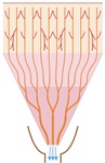 The whole collecting duct
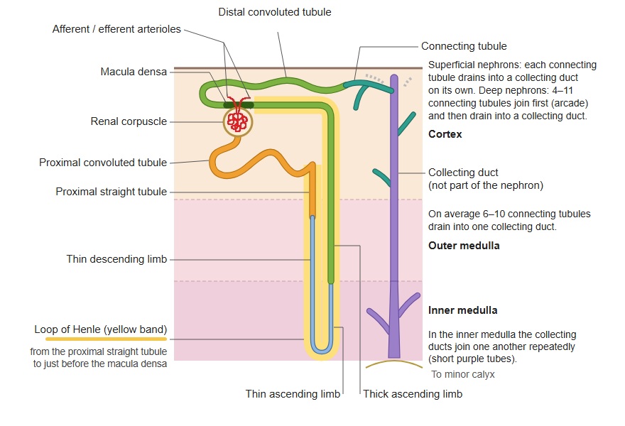
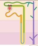 Nephron and collecting duct
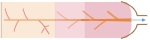 Collecting duct (schematic)
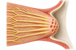 Renal papilla
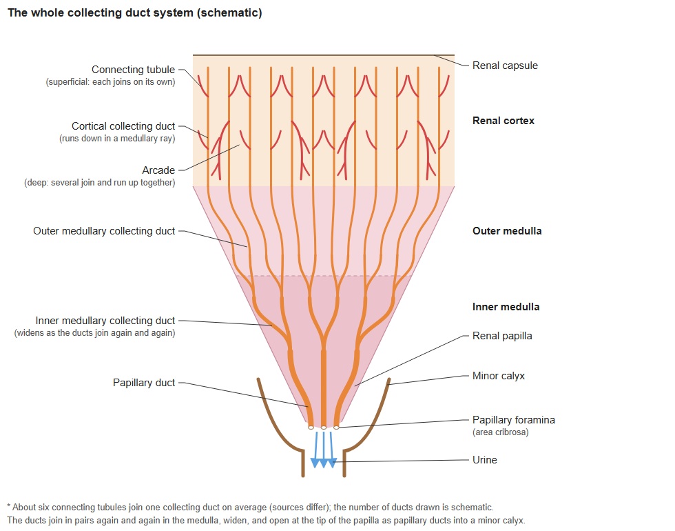
- Renal cortex
- Renal corpuscle : glomerulus and Bowman's capsule.
- Proximal tubule : takes back water and nutrients from the primary urine.
- Distal tubule : adjusts electrolytes and water.
- Cortical collecting duct : collects primary urine from several connecting tubules.
- Renal medulla
- Renal pyramid : the cone-shaped tissue that forms the medulla; it contains:
- Loop of Henle : part of the countercurrent system that lets the kidney concentrate urine.
- Medullary collecting duct : carries primary urine to the renal papilla.
- Papillary duct : opens at the tip of the papilla; its contents leave into a minor calyx as urine.
- Renal pyramid : the cone-shaped tissue that forms the medulla; it contains:
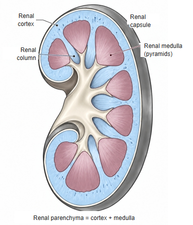
renal medulla (click to enlarge)
Reference websites
[Wikipedia]
· Collecting duct system, Wikipedia.
· Renal papilla, Wikipedia.
[Websites with illustrations]
· Tubules and collecting system (Digital Histology)
· Tubules and collecting system 17 (Digital Histology)
· Microscopic anatomy, plate 12.240 (Anatomy Atlases)
Related terms
- Proximal tubule
- Straight tubule
- Distal tubule
- Cortical collecting duct
- Medullary collecting duct
- Kidney
- Renal parenchyma
- Renal pelvis
- Ureter
- Urinary system
References and sources
- Further reading: Collecting duct system, Wikipedia.
- Further reading: Renal papilla, Wikipedia.
- Federative International Programme for Anatomical Terminology (FIPAT). Terminologia Anatomica, 2nd ed. (TA2), 2019.