Foot process
Also called the pedicel
Overview
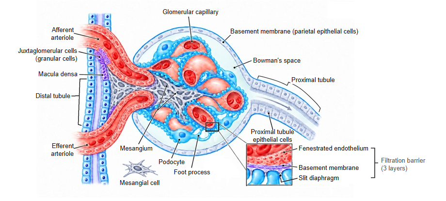
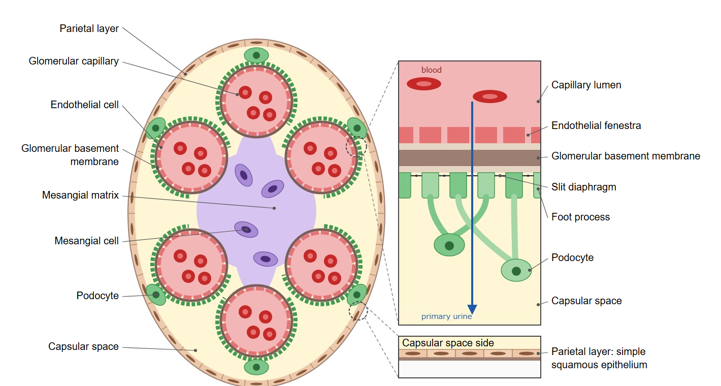
· Foot processes are the slender processes that branch from the primary processes of a podocyte and attach to the outer surface of the glomerular basement membrane. They are also called pedicels.
· Podocytes form the visceral layer of the glomerular capsule (Bowman's capsule), and their name comes from these foot-like processes, which each cell has in very large numbers.
· The foot processes of neighbouring podocytes interdigitate like interlocked fingers and wrap the glomerular capillaries from outside. The narrow gaps between them are the filtration slits, and across each gap, close to the glomerular basement membrane, stretches the slit diaphragm.
· Inside each foot process is a cytoskeleton made mainly of actin, which keeps its shape and supports its attachment to the basement membrane. The surface is covered by a negatively charged coat, which helps hold back negatively charged proteins such as albumin.
· When podocytes are injured, the foot processes retract and spread out flat, and the interdigitating pattern is lost (foot process effacement). This is seen in minimal change nephrotic syndrome and other diseases; protein then leaks into the primary urine, causing proteinuria.
Reference websites
[Wikipedia]
· Podocyte, Wikipedia.
· Foot process effacement, Wikipedia.
[Websites with illustrations]
· Podocytes (OpenStax illustration and micrograph) (Wikimedia Commons)

· Podocyte morphology (Wikimedia Commons)

· Glomerulus in the scanning electron microscope (Wikimedia Commons)

· Renal corpuscle (electron micrograph) (Histology Guide)
Related terms
- Podocyte
- Primary process
- Filtration slit
- Slit diaphragm
- Glomerular basement membrane
- Visceral layer of glomerular capsule
- Glomerular capillary
References and sources
- Further reading: Podocyte, Wikipedia.
- Further reading: Foot process effacement, Wikipedia.
- Federative International Programme for Anatomical Terminology (FIPAT). Terminologia Anatomica, 2nd ed. (TA2), 2019.