Dilator pupillae (Latin: musculus dilatator pupillae)
Also called: dilator pupillae muscle; iris dilator muscle; pupillary dilator
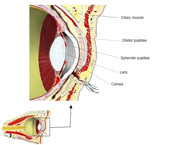
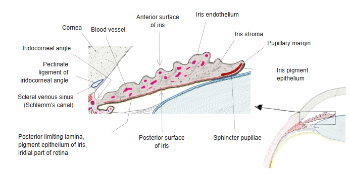
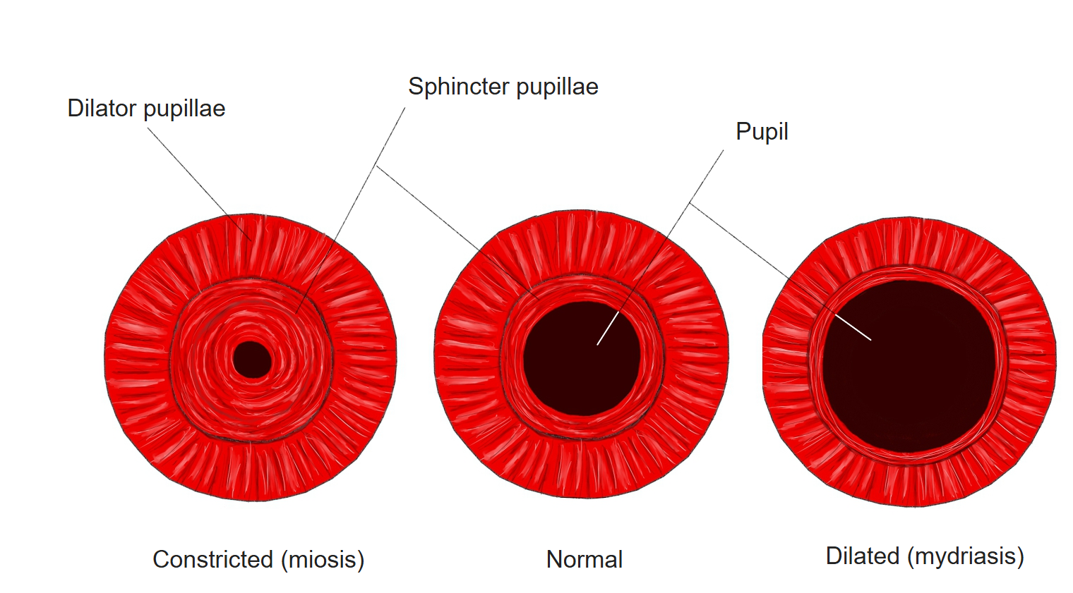
· Its fibres run radially, like the spokes of a wheel, from the outer edge of the iris toward the centre of the pupil; they lie just in front of the pigmented epithelium at the back of the iris.
· When it contracts, the iris is pulled outward and the pupil becomes larger (mydriasis), letting more light reach the retina, as in dim light or when sympathetic activity is high.
· It is smooth muscle, made of modified myoepithelial cells that develop from the neuroectoderm of the optic cup.
· It is the antagonist of the sphincter pupillae; the two together are called the muscles of the iris.
· Nerve: sympathetic fibres from the superior cervical ganglion; they reach the eye with the internal carotid artery and the nasociliary nerve and enter it in the long ciliary nerves and the short ciliary nerves (passing through the ciliary ganglion without a synapse). Noradrenaline acts on alpha-1 receptors.
muscles
- Ciliary muscle : surrounds the lens and adjusts its thickness.
- Sphincter pupillae : runs in a ring in the iris and makes the pupil smaller. With the dilator pupillae it forms the muscles of the iris.
- Dilator pupillae : runs radially in the outer part of the iris and makes the pupil larger.
Nerve fibres passing the ciliary ganglion
Some sources divide the muscles of the eye into intra-ocular and extra-ocular muscles. Others use 'ocular muscles' only for the muscles that move the eyeball and leave out the intra-ocular muscles, probably because these are smooth muscle.
In that narrower sense the ocular muscles are the following; some authors also leave out the levator palpebrae superioris at the bottom of the list.
- Recti : superior rectus, inferior rectus, medial rectus, lateral rectus
- Obliques : superior oblique, inferior oblique (the recti and obliques move the eyeball)
- Levator palpebrae superioris : raises the upper eyelid.
- Nerve supply : oculomotor nerve (superior, inferior and medial rectus, inferior oblique, levator palpebrae superioris), trochlear nerve (superior oblique), abducens nerve (lateral rectus).
The following classification divides the ocular muscles broadly into the extra-ocular muscles (striated muscle) and the intra-ocular muscles (smooth muscle).
muscles (9)
- Extra-ocular muscles (6)
- Recti : superior rectus, inferior rectus, medial rectus, lateral rectus
- Obliques : superior oblique, inferior oblique
- Intra-ocular muscles (3) : ciliary muscle, sphincter pupillae, dilator pupillae
- Other muscles in the orbit : levator palpebrae superioris, orbitalis, superior tarsal muscle, inferior tarsal muscle
Reference websites
[Wikipedia]
· Iris dilator muscle, Wikipedia.
[Websites with illustrations]
· Iris: anatomy and function (Kenhub)
Related terms
- Muscles of iris
- Intra-ocular muscles
- Ciliary muscle
- Pigmented epithelium of iris
- Stroma of iris
- Myoepithelial cell
- Smooth muscle
References and sources
- Further reading: Iris dilator muscle, Wikipedia.
- Federative International Programme for Anatomical Terminology (FIPAT). Terminologia Anatomica, 2nd ed. (TA2), 2019.