Suspensory ligament of ovary
Also called the infundibulopelvic ligament.
· One of the ligaments that fix the ovary; also called the infundibulopelvic ligament (IP ligament), because it reaches the pelvic wall from the infundibulum of the uterine tube.
of the
ovary
- Proper ligament of ovary : joins the uterine (lower) end of the ovary to the lateral angle of the uterus, just below the uterine tube; a cord of connective tissue and smooth muscle
- Suspensory ligament of ovary : joins the tubal (upper) end of the ovary to the lateral wall of the pelvis; a peritoneal fold with connective tissue and smooth muscle
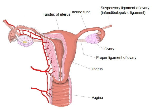
The ovary is held in position by two bands: the proper ligament of the ovary and the suspensory ligament of the ovary. The proper ligament is a short, round cord of connective tissue and smooth muscle that lies between the anterior and posterior layers of the broad ligament and joins the lateral angle of the uterus to the uterine end of the ovary. The suspensory ligament likewise contains connective tissue and smooth muscle; it stretches from the tubal end of the ovary to the lateral pelvic wall and carries the vessels and nerves of the ovary.
It is a fold of peritoneum that extends from the ovary to the wall of the pelvis and passes upwards over the external iliac vessels (external iliac artery and vein). Some sources treat it as part of the broad ligament of the uterus, others as the lateral end of that ligament. Although it is called a ligament, it gives the ovary little mechanical support; it matters mainly as a landmark and as the route of the ovarian vessels.
Structure
It contains the ovarian artery, the ovarian vein, the ovarian plexus of nerves and the lymphatic vessels of the ovary.
Medially it is continuous with the broad ligament, which is attached to the upper pole of the ovary and the infundibulum of the uterine tube; laterally it becomes the parietal peritoneum of the pelvic wall. It is therefore one continuous peritoneal fold whose parts carry different names. Near the pelvic brim the ureter runs close to it, just medial and posterior, so the ureter must be identified when the ovarian vessels are tied in this ligament during surgery.
Reference websites
[Websites with illustrations]
· Arterial supply of the ovary, with the suspensory ligament (Earth's Lab)
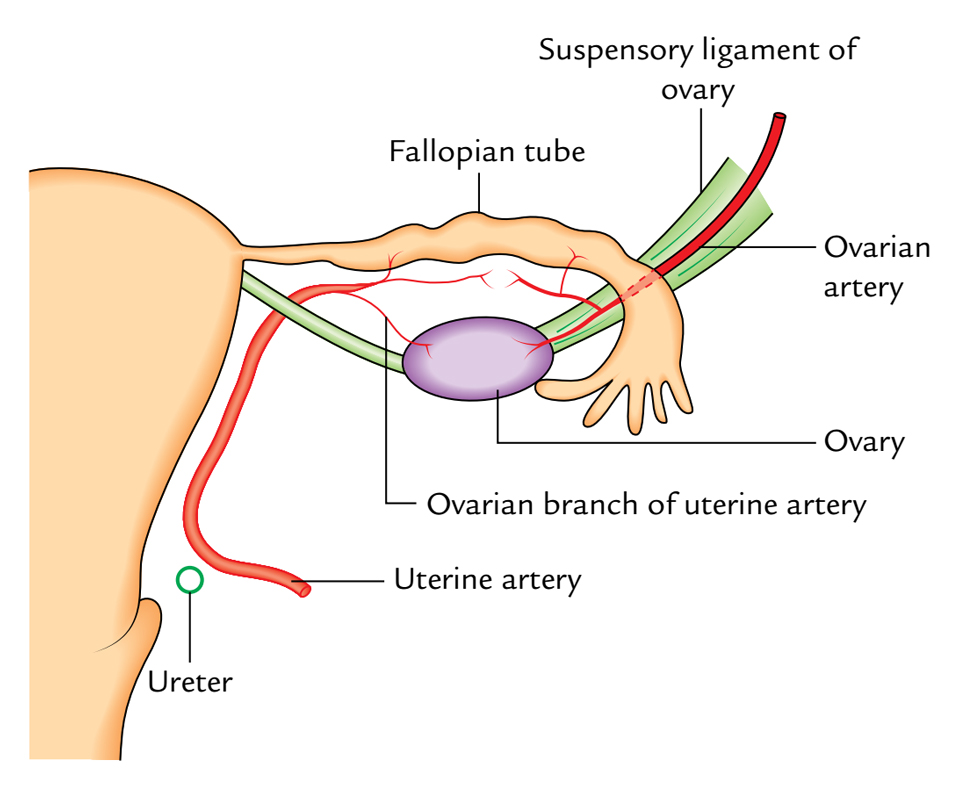
· Ovary, uterine tube and their ligaments (Earth's Lab)
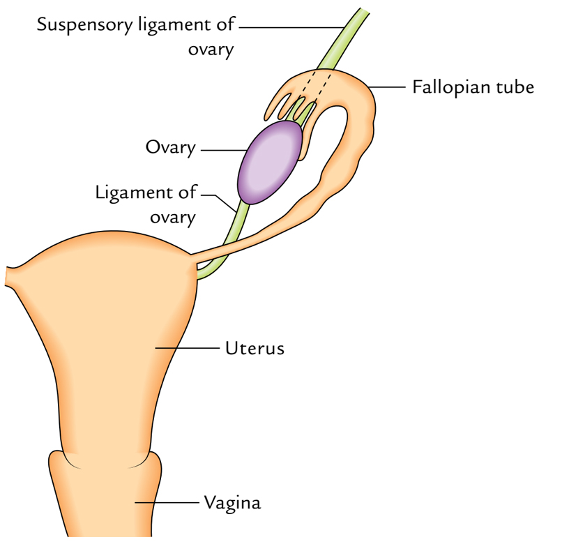
· The ovaries (TeachMeAnatomy)
· Suspensory ligament of the ovary (Radiopaedia)
Related terms
- Ovary
- Uterus
- Uterine tube
- Peritoneum
- Peritoneal folds
- Ovarian artery
- Ovarian vein
- Ovarian plexus
- Ureter
- Lesser pelvis
- Ligament
References and sources
- Suspensory ligament of ovary, Wikipedia.
- FIPAT. Terminologia Anatomica, 2nd ed. (TA2). Federative International Programme for Anatomical Terminology, 2019: suspensory ligament of ovary (ligamentum suspensorium ovarii), TA2 3805.