← Multi-nerve illustrations: Spinal nerves
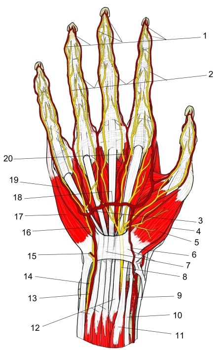
| No. | Name | Page |
|---|---|---|
| 1 | Proper palmar digital arteries | Detail |
| 2 | Proper palmar digital nerves | — |
| 3 | Common palmar digital nerves | Detail |
| 4 | Opponens pollicis | Detail |
| 5 | Abductor pollicis brevis | Detail |
| 6 | Superficial palmar branch (radial artery) | Detail |
| 7 | Flexor retinaculum | — |
| 8 | Median nerve | Detail |
| 9 | Radial artery | Detail |
| 10 | Flexor carpi radialis (tendon) | Detail |
| 11 | Flexor pollicis longus | Detail |
| 12 | Flexor digitorum superficialis | Detail |
| 13 | Ulnar artery | Detail |
| 14 | Ulnar nerve | Detail |
| 15 | Deep branch (ulnar nerve and ulnar artery) | — |
| 16 | Superficial branch (ulnar nerve) | Detail |
| 17 | Abductor digiti minimi (hand) | Detail |
| 18 | Superficial palmar arch | Detail |
| 19 | Flexor digiti minimi brevis (hand) | Detail |
| 20 | Common palmar digital arteries | Detail |
More illustrations: Spinal nerves
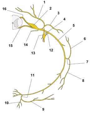 Typical spinal nerve: anterior and posterior rami (schematic)
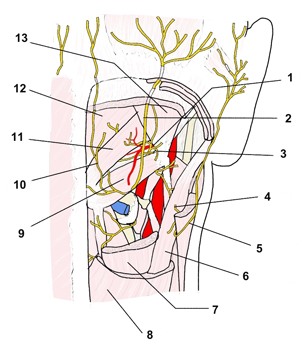 Cervical nerves, suboccipital region (posterior view)
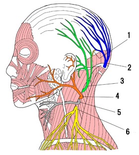 Cervical plexus, cutaneous branches (lateral view)
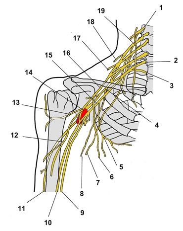 Cervical and brachial plexuses, right side (anterior view)
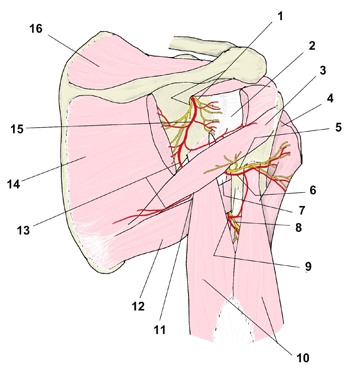 Scapular region, nerves and arteries (posterior view)
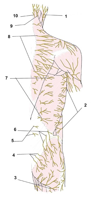 Nerves of the back, cutaneous branches (posterior view)
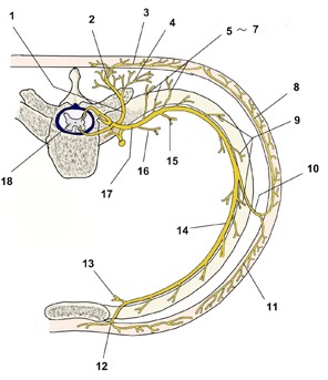 Thoracic spinal nerve and intercostal nerve branches (transverse schematic)
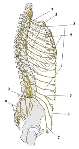 Intercostal nerves and nerves of the trunk wall (lateral view)
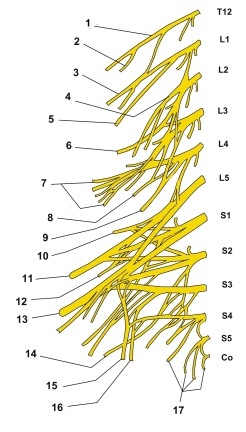 Lumbosacral plexus (schematic)
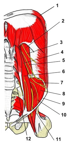 Lumbar plexus on the posterior abdominal wall (anterior view)
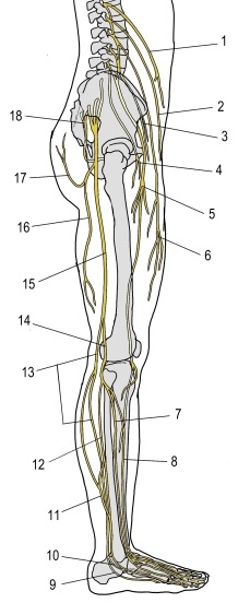 Lumbar and sacral plexuses, right lower limb (lateral view)
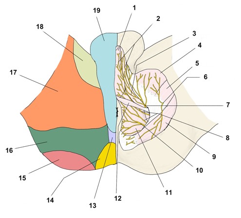 Pudendal plexus: nerves and cutaneous territories of the male perineum (inferior view)
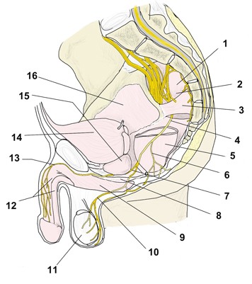 Pudendal plexus (median section)
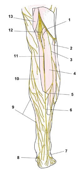 Nerves of the leg (posterior view)
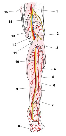 Leg, nerves and arteries (posterior view)
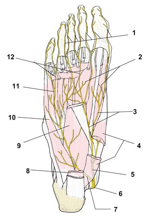 Nerves of the sole of the foot
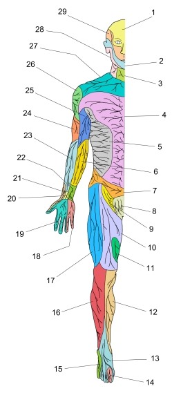 Cutaneous nerve distribution (anterior view)
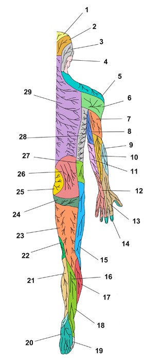 Cutaneous nerve distribution (posterior view)