Latin: sternum · the breastbone
Overview
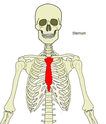
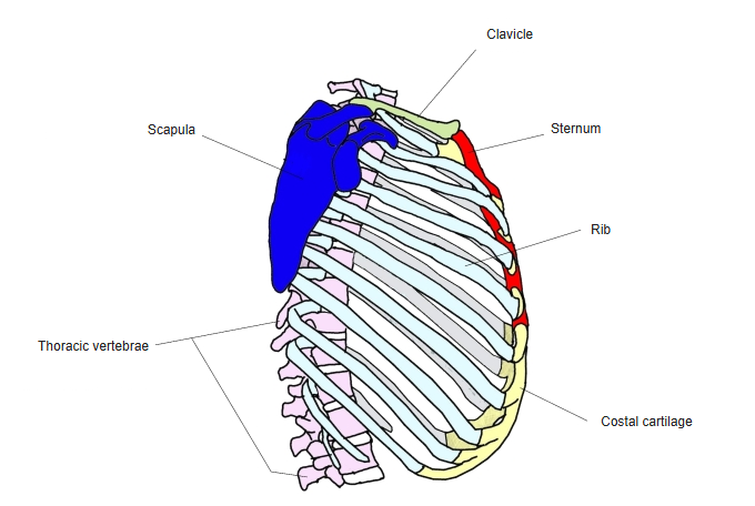
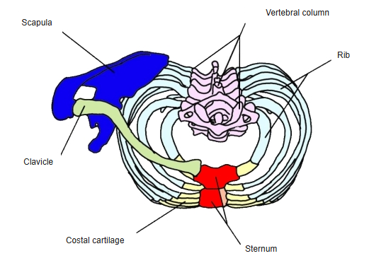
- One of the bones of the thorax, lying in the midline of its front wall.
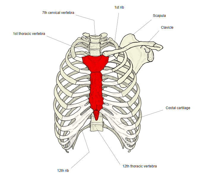
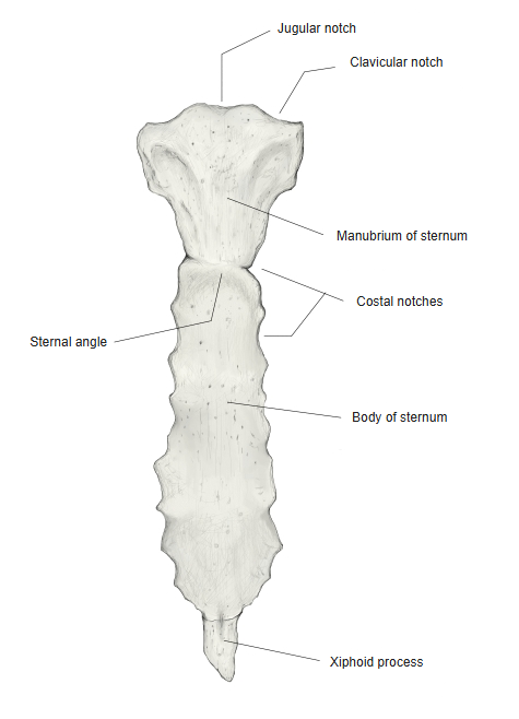
- A long, flat bone with three parts, from above downwards: the manubrium, the body and the xiphoid process.
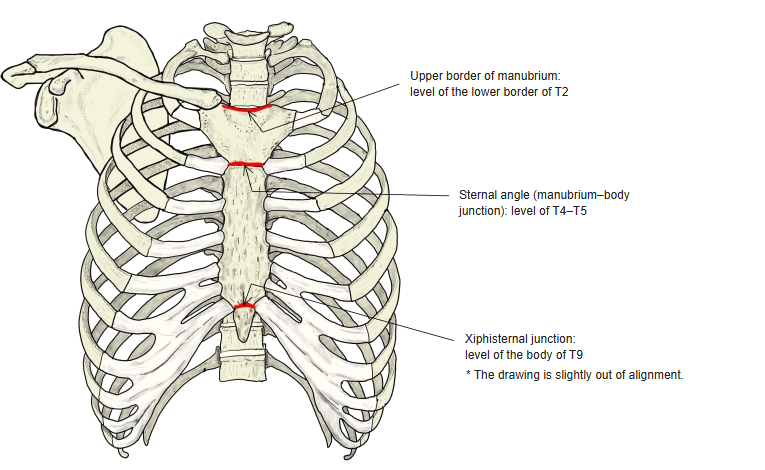
- Levels: the upper border of the manubrium is at the lower border of T2; the sternal angle (manubrium–body junction) at T4–T5; the junction of the body and xiphoid process at the body of T9.
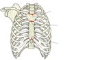
Vertebral levels of the sternum
- Bones of the trunk
- Vertebral column
- Cervical vertebrae (7)
- Thoracic vertebrae (12)
- Lumbar vertebrae (5)
- Sacrum (1)
- Coccyx (1)
- Thoracic cage
- Sternum (1)
- Ribs (24)
- Vertebral column
Illustrations
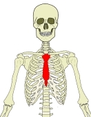
Position of the sternum 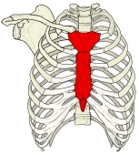
Thorax (anterior) 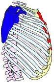
Thorax (lateral) 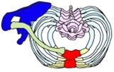
Thorax (horizontal section) 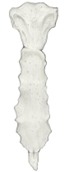
Sternum (anterior) 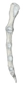
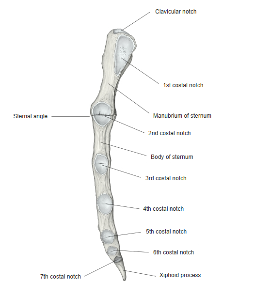
Sternum (left lateral)
Articulations
| # | Bone | Joint |
|---|---|---|
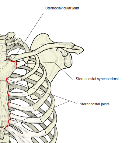
| 1 | Clavicle (sternal facet) ⇔ sternum (clavicular notch) | Sternoclavicular joint |
| 2 | Costal cartilages 2–7 ⇔ sternum (costal notches) | Sternocostal joints |
| 3 | 1st costal cartilage ⇔ 1st costal notch | Sternocostal synchondrosis |
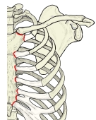
Joints of the sternum
Ligaments
The following ligaments attach to the sternum. The list is intended to be comprehensive but may omit minor ligaments.
| # | Ligament | Connects |
|---|---|---|
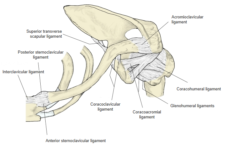
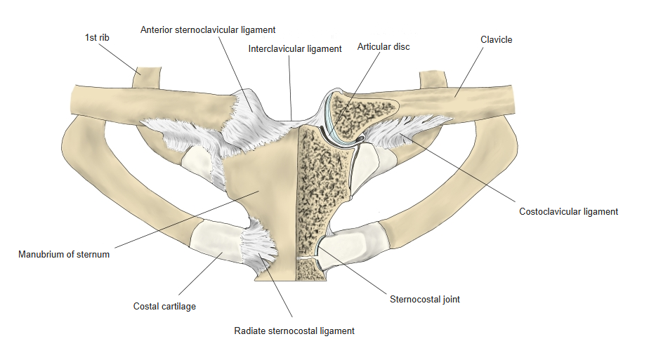
| 1 | Posterior sternoclavicular ligament | Sternoclavicular joint: back of the sternal end of the clavicle and the manubrium |
| 2 | Anterior sternoclavicular ligament | Sternoclavicular joint: front of the sternal end of the clavicle and the manubrium |
| 3 | Interclavicular ligament | Between the upper parts of the sternal ends of both clavicles, attached also to the upper border of the manubrium |
| 4 | Radiate sternocostal ligaments | Sternocostal joints: ends of the costal cartilages – sternum |
| 5 | Intra-articular sternocostal ligament | Divides the cavity of the 2nd sternocostal joint in two |
| 6 | Costoxiphoid ligaments | 6th and 7th costal cartilages – xiphoid process |
Ligaments of the clavicle and sternum (anterior) 
Sternoclavicular and sternocostal joints
Muscle attachments
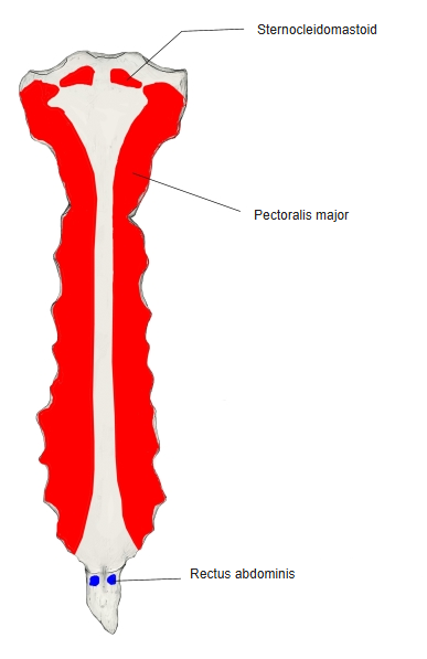
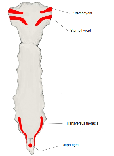
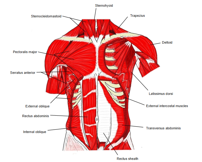
- Origin of: Sternocleidomastoid, Pectoralis major, Sternohyoid, Sternothyroid, Transversus thoracis, Diaphragm
- Insertion of: Rectus abdominis
Sternocleidomastoid: upper part of the front of the manubrium · pectoralis major: front, from the manubrium to the lower body · sternohyoid and sternothyroid: back of the manubrium · transversus thoracis: back of the lower body and of the xiphoid process · diaphragm: back of the xiphoid process · rectus abdominis: front of the xiphoid process.
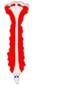
Muscle attachments (anterior) 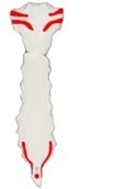
Muscle attachments (posterior) 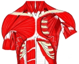
Muscles of the chest (anterior)
Development
- The body of the sternum forms from four pieces (sternebrae) joined by cartilage; in the adult they fuse, and the lines of fusion remain as transverse ridges on the body.
- The cartilaginous joints between manubrium and body (manubriosternal joint) and between body and xiphoid process (xiphisternal joint) last longer, but most ossify in old age.
Development of the sternum
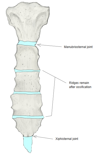
Variation
- A congenital hole in the body or xiphoid process is called a sternal foramen; failure of the two halves of the sternum to fuse is called a sternal cleft.
Etymology
- From Greek sternon, “chest”.
References and sources
- Further reading: Sternum, Wikipedia.