Latin: atlas · first cervical vertebra (C1)
Overview
- The first cervical vertebra (C1) and the uppermost vertebra of the vertebral column; it carries the skull, and is named after the Titan Atlas who held up the heavens.
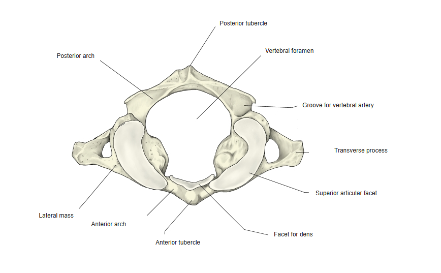
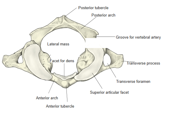
- Unlike the other cervical vertebrae it has no vertebral body and no spinous process: seen from above it is a ring made of an anterior arch, a posterior arch and two lateral masses.
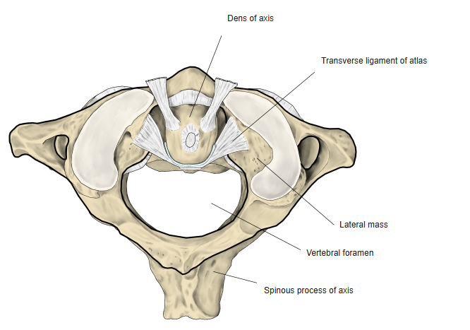
- The transverse ligament of the atlas divides the vertebral foramen into two parts: the front part receives the dens of the axis, and the larger back part is the true vertebral foramen for the spinal cord.
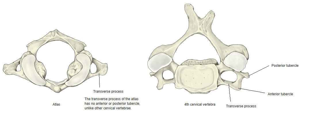
- The atlas has anterior and posterior tubercles, but they lie at the middle of the anterior and posterior arches – not at the tips of the transverse processes as in other cervical vertebrae.
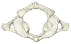
Atlas (superior view) 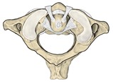
Atlas with the dens of the axis
- Bones of the trunk
- Vertebral column
- Cervical vertebrae (7)
- Thoracic vertebrae (12)
- Lumbar vertebrae (5)
- Sacrum (1)
- Coccyx (1)
- Thoracic cage
- Sternum (1)
- Ribs (24)
- Vertebral column
Illustrations
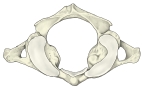
Atlas (from above) 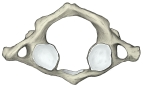
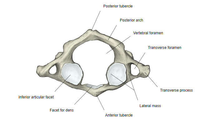
Atlas (from below) 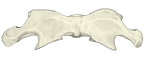
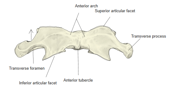
Atlas (anterior) 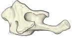
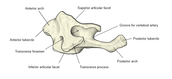
Atlas (left lateral) 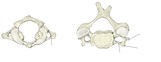
Transverse process: atlas and 4th cervical vertebra 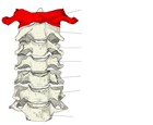
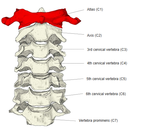
Cervical vertebrae (anterior), atlas highlighted 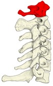
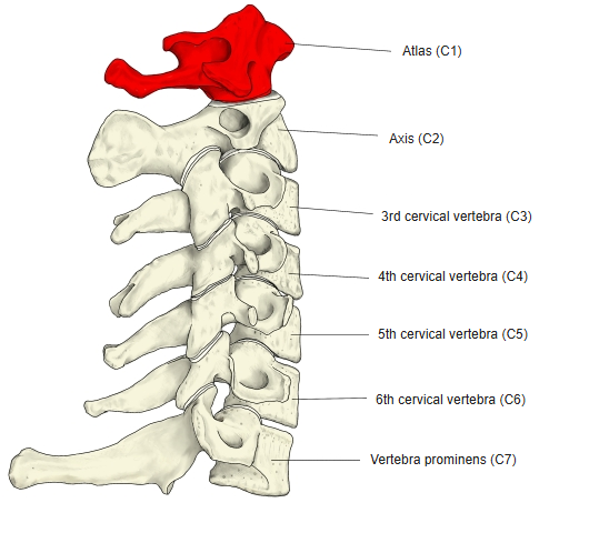
Cervical vertebrae (left lateral), atlas highlighted 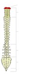
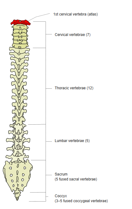
Vertebral column, atlas highlighted
Articulations
| # | Bone | Joint |
|---|---|---|
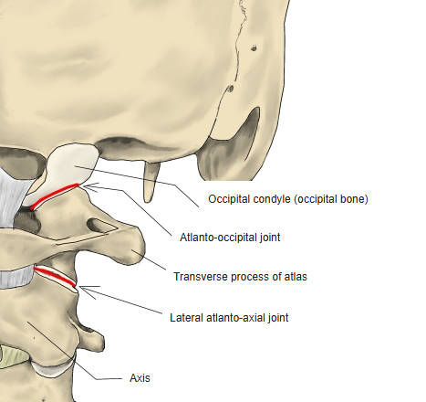
| 1 | Occipital bone | Atlanto-occipital joint (superior articular facets – occipital condyles) |
| 2 | Axis | Median atlanto-axial joint (facet for dens – dens of axis) |
| 3 | Axis | Lateral atlanto-axial joint (inferior articular facets – superior articular facets of axis) |
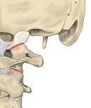
Joints of the atlas
Ligaments
The following ligaments attach to the atlas. The list is intended to be comprehensive but may omit minor ligaments.
| # | Ligament | Connects |
|---|---|---|
| 1 | Anterior longitudinal ligament | Occipital bone (pharyngeal tubercle) – front of the sacrum; attaches to the anterior tubercle of the atlas |
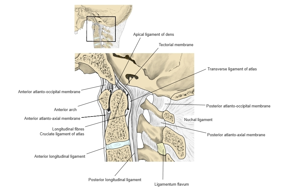
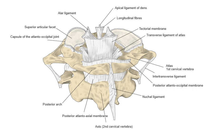
| 2 | Transverse ligament of atlas (part of the cruciate ligament) | Between the lateral masses of the atlas, behind the dens |
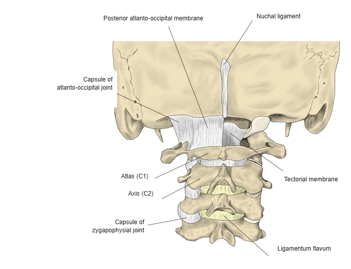
| 3 | Nuchal ligament | Occipital bone – posterior tubercle of the atlas and spinous processes of the cervical vertebrae |
| 4 | Intertransverse ligaments | Between the transverse processes |
| 5 | Lateral atlanto-occipital ligament | Occipital bone (jugular process) – transverse process of the atlas |
| 6 | Anterior atlanto-occipital membrane | Front margin of the foramen magnum – anterior arch of the atlas |
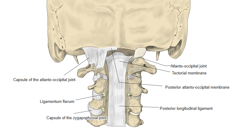
| 7 | Posterior atlanto-occipital membrane | Back margin of the foramen magnum – posterior arch of the atlas |
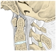
Ligaments of the craniovertebral junction (median section) 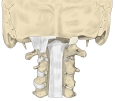
Ligaments (posterior, vertebral canal opened) 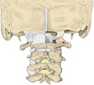
Ligaments of the craniovertebral junction 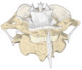
Ligaments of the atlas and axis (posterior)
Muscle attachments
- Origin of: Rectus capitis posterior minor, Obliquus capitis superior, Rectus capitis anterior, Rectus capitis lateralis, Levator scapulae
- Insertion of: Longus colli, Obliquus capitis inferior, Splenius cervicis, Intertransversarii
Rectus capitis posterior minor arises from the posterior tubercle; rectus capitis anterior from the lateral mass; obliquus capitis superior, rectus capitis lateralis and levator scapulae from the transverse process. Longus colli inserts on the anterior tubercle; obliquus capitis inferior and splenius cervicis insert on the transverse process.
Parts
- Lateral mass – the thick part on each side where the anterior and posterior arches meet; it carries the articular facets.
- Superior articular facet – the concave, kidney-shaped facet on the upper surface of the lateral mass; articulates with the occipital condyle of the occipital bone.
- Inferior articular facet – the flat, roughly circular facet on the lower surface of the lateral mass; articulates with the superior articular facet of the axis.
- Anterior arch – the short front arch of the ring.
- Anterior tubercle – a small elevation at the middle of the front of the anterior arch; longus colli attaches here.
- Facet for dens – a small, smooth facet on the back of the anterior arch; articulates with the dens of the axis.
- Posterior arch – the longer back arch of the ring.
- Posterior tubercle – a small elevation at the middle of the posterior arch, in place of a spinous process; rectus capitis posterior minor arises here.
- Groove for vertebral artery – a shallow groove on the upper surface of the posterior arch, behind the lateral mass, for the vertebral artery (and the suboccipital nerve).
- Transverse process – projects further laterally than in the other cervical vertebrae and contains the transverse foramen; it has no anterior or posterior tubercle.
- Vertebral foramen – very large, enclosed by the two arches and the lateral masses.
References and sources
- Further reading: Atlas (anatomy), Wikipedia.