Foramen magnum (Latin: foramen magnum)
The large opening in the occipital bone, bounded by its basilar part in front, its lateral parts at the sides and its squamous part behind. It joins the cranial cavity to the vertebral canal and transmits several nerves, vessels and other structures.
· It lies in the floor of the posterior cranial fossa and is longer from front to back than from side to side.
· The occipital condyles lie along the sides of its front half, and the hypoglossal canals pierce the bone above them.
· The midpoint of its front margin is the basion; the midpoint of its back margin is the opisthion.
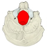 Occipital bone (inferior)
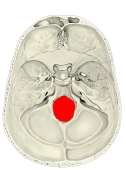 Cranial base (internal surface)
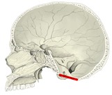 Skull (sagittal section)
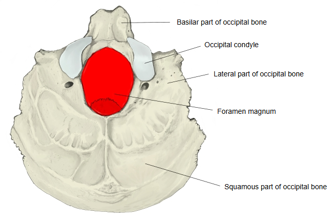
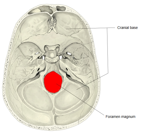
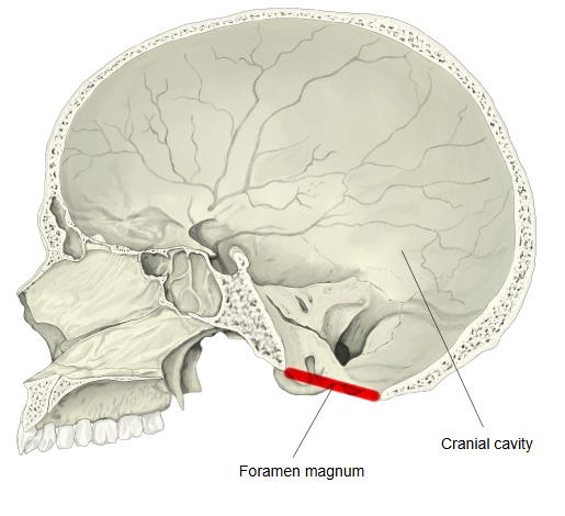
More details
It is an oval opening, longer from front to back, and the nerves and vessels below pass through it. (Note: sources differ somewhat on what passes through the foramen magnum.)
| Category | Name | Illustration | |
|---|---|---|---|
| Nervous system | Medulla oblongata | its lower end, continuing into the spinal cord | 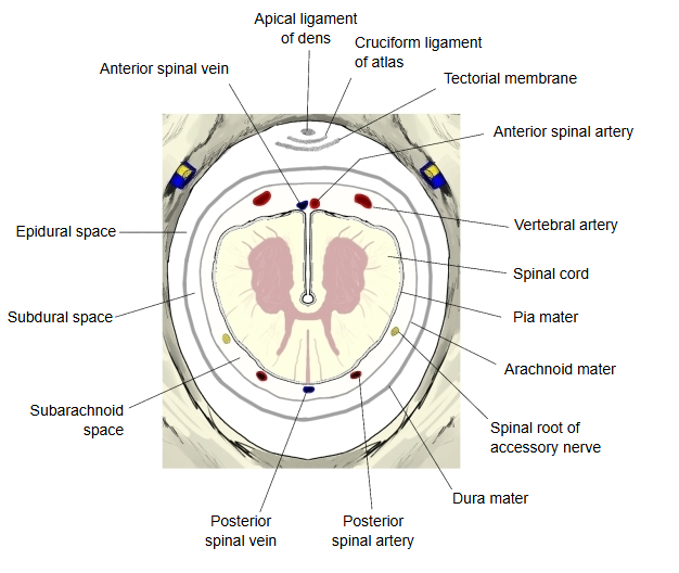 |
| Accessory nerve, spinal root | runs up from the upper cervical cord into the skull | ||
| Arteries | Vertebral artery | right and left; they join above the opening to form the basilar artery | |
| Anterior spinal artery | formed by branches of the two vertebral arteries | ||
| Posterior spinal arteries | from the vertebral artery or the posterior inferior cerebellar artery | ||
| Veins | Anterior spinal veins | join the venous plexuses around the opening | |
| Posterior spinal veins | join the venous plexuses around the opening | ||
| Ligaments | Cruciform ligament of atlas | its upper band runs up to the front margin | |
| Apical ligament of dens | from the tip of the dens to the front margin | ||
| Others | Tectorial membrane | runs up to the front margin, behind the ligaments above | |
| Meninges (dura mater, arachnoid mater, pia mater) | around the medulla and spinal cord; continuous from the skull into the vertebral canal | ||
Note: some sources also list the cerebellar tonsils, which normally lie just above the opening; they are not included here. Some sources, on the other hand, give no veins at all; here the anterior and posterior spinal veins are included.
Reference websites
[Wikipedia]
· Foramen magnum, Wikipedia.
[Websites with illustrations]
· Skull from below, foramen magnum (Wikimedia Commons)
· Foramen magnum (inferior view) (Wikimedia Commons)
· The occipital bone (TeachMeAnatomy)
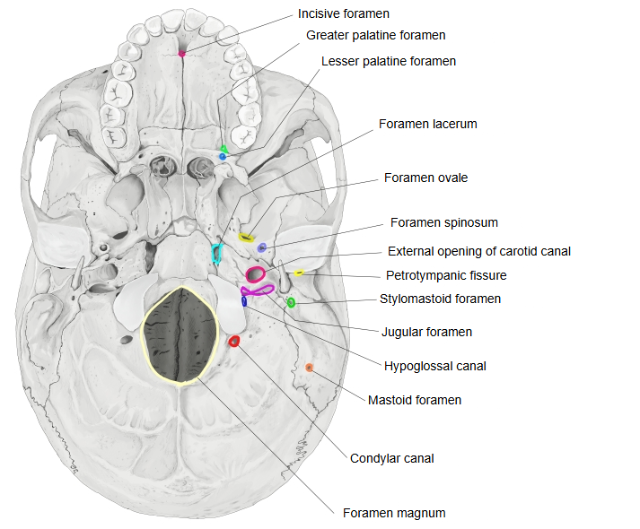
 Foramina of the external cranial base
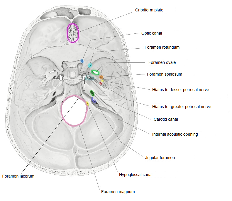
 Foramina of the internal cranial base


Where it is found
Related terms
- Occipital condyle
- Hypoglossal canal
- Clivus
- Jugular foramen
- Occipital squama
- Atlanto-occipital joint
- Occipital bone
References and sources
- Further reading: Foramen magnum, Wikipedia.
- Further reading: Occipital bone, Wikipedia.
- Federative International Programme for Anatomical Terminology (FIPAT). Terminologia Anatomica, 2nd ed. (TA2), 2019.