Latin: os palatinum
Overview
- A paired, L-shaped bone at the back of the nasal cavity, lying between the maxilla in front and the pterygoid process of the sphenoid behind.
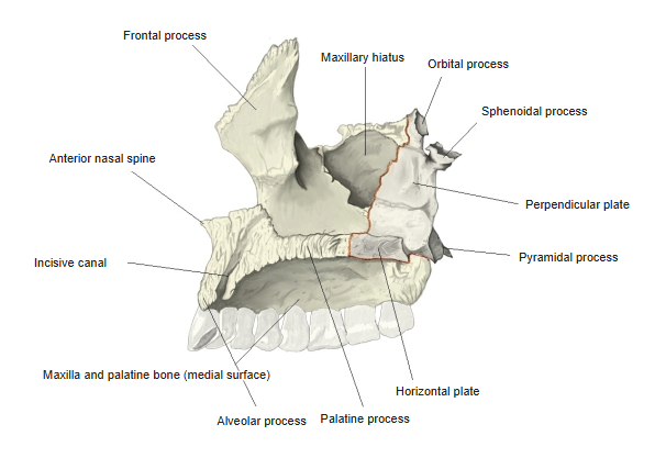
- It has two plates – the horizontal plate, forming the back of the hard palate, and the perpendicular plate, forming part of the lateral wall of the nasal cavity – and three processes: the pyramidal, orbital and sphenoidal processes.
- It contributes to the floor and lateral wall of the nasal cavity, the roof of the mouth, the floor of the orbit and the pterygopalatine fossa.
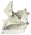
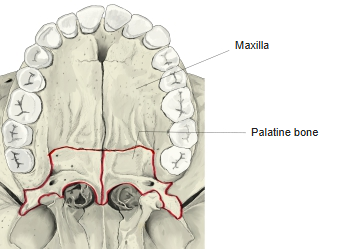
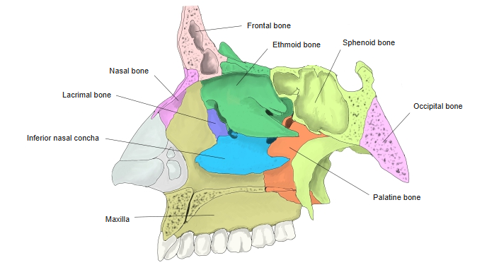
Maxilla and palatine bone (medial) 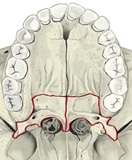
Hard palate (from below) 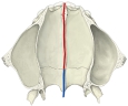
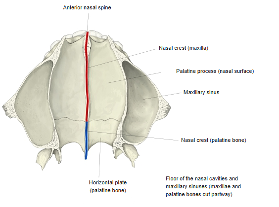
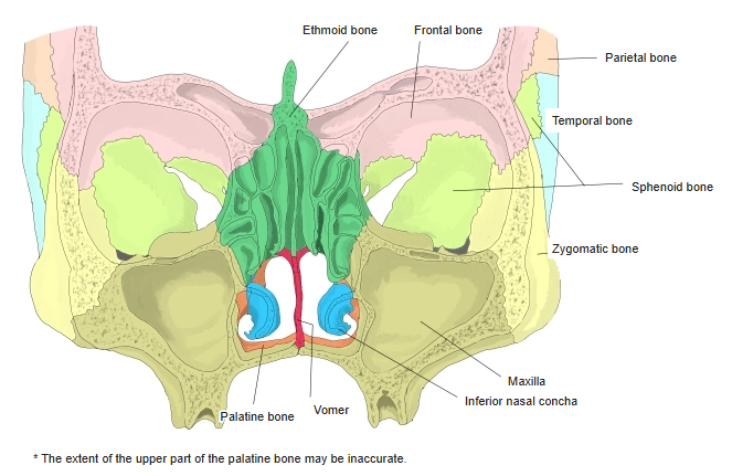
Floor of the nasal cavity (from above)
- Bones of the skull
- Neurocranium (cranial bones)
- Frontal bone
- Parietal bone (2)
- Occipital bone
- Temporal bone (2)
- Sphenoid bone
- Ethmoid bone
- Viscerocranium (facial bones)
- Nasal bone (2)
- Vomer
- Lacrimal bone (2)
- Inferior nasal concha (2)
- Maxilla (2)
- Zygomatic bone (2)
- Palatine bone (2)
- Mandible
- Hyoid bone
- Neurocranium (cranial bones)
Illustrations
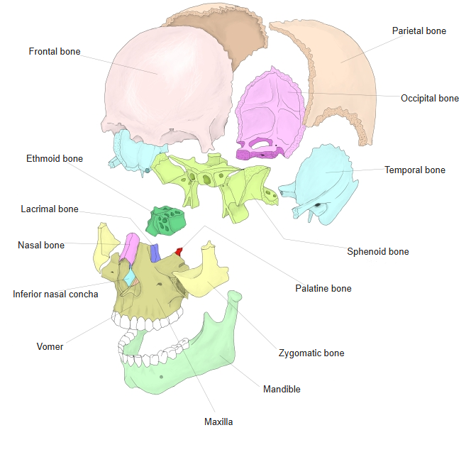
Bones of the skull (separated)
Articulations
| # | Bone | Joint |
|---|---|---|
| 1 | Maxilla | Transverse palatine suture; nasal surface |
| 2 | Opposite palatine bone | Median palatine suture |
| 3 | Sphenoid bone | Sphenoidal process and pyramidal process |
| 4 | Ethmoid bone | Orbital process and ethmoidal crest |
| 5 | Inferior nasal concha | Conchal crest |
| 6 | Vomer | Nasal crest |
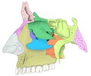
Lateral wall of the nasal cavity 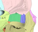
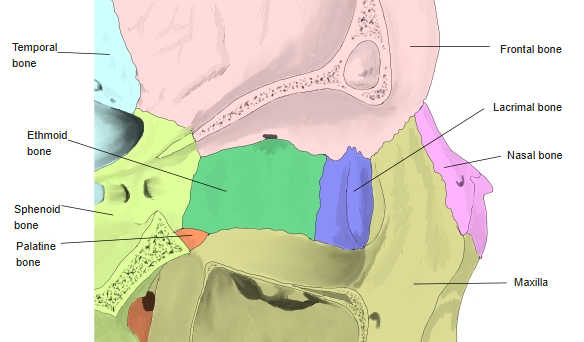
Medial wall of the orbit 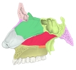
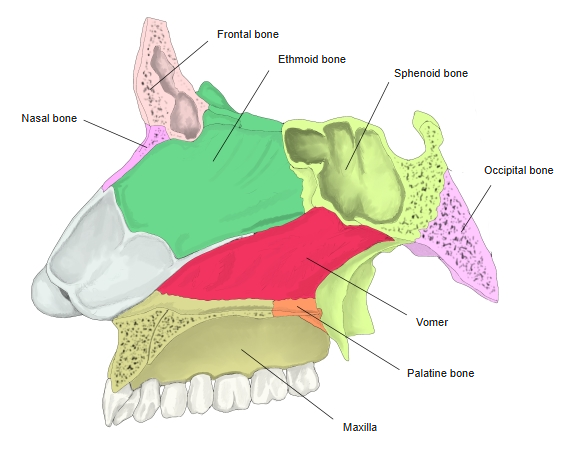
Sagittal section of the nasal cavity 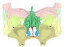
Coronal section through the nasal cavity
Muscle attachments
- Origin of: Medial pterygoid (deep head, from the pyramidal process), Tensor veli palatini (via the palatine aponeurosis), Musculus uvulae (from the posterior nasal spine), Superior pharyngeal constrictor (in part)
- Insertion of: none
The palatine aponeurosis attaches to the posterior border of the horizontal plate.
Plates and processes
- Horizontal plate – meets the palatine process of the maxilla in front at the transverse palatine suture and the opposite plate at the median palatine suture; its free posterior border ends medially in the posterior nasal spine. Its upper surface rises as the nasal crest, which meets the vomer.
- Perpendicular plate – a thin plate on the nasal surface of the maxilla, partly covering the maxillary hiatus; it carries the conchal crest for the inferior nasal concha and the ethmoidal crest for the middle concha. The greater palatine groove on its lateral side forms the greater palatine canal with the maxilla.
- Pyramidal process – projects backwards and laterally from the junction of the plates, fitting between the plates of the pterygoid process; it carries the lesser palatine foramina.
- Orbital process – at the top of the perpendicular plate, forming a small part of the floor of the orbit.
- Sphenoidal process – behind the orbital process, meeting the body of the sphenoid; the notch between the two processes is closed by the sphenoid to form the sphenopalatine foramen.
References and sources
- Further reading: Palatine bone, Wikipedia.