Sphenopalatine foramen (Latin: foramen sphenopalatinum)
Nerves and vessels passing through the sphenopalatine foramen
Textbooks name these structures in slightly different ways.
A standard textbook: the posterior superior nasal nerves (from the pterygopalatine ganglion) and the nasopalatine artery and vein.
Prometheus atlas: the lateral and medial superior posterior nasal branches of the maxillary nerve (lateral, medial), and the sphenopalatine artery with its accompanying vein.
Wikipedia: the posterior superior lateral nasal nerves, the nasopalatine nerve, and the sphenopalatine artery and vein.
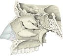 Right nasal cavity (lateral wall)
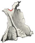 Palatine bone (maxillary surface)
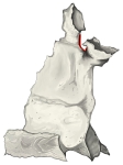 Palatine bone (nasal surface)
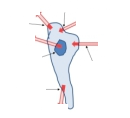 Pterygopalatine fossa (diagram)
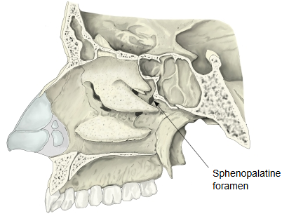
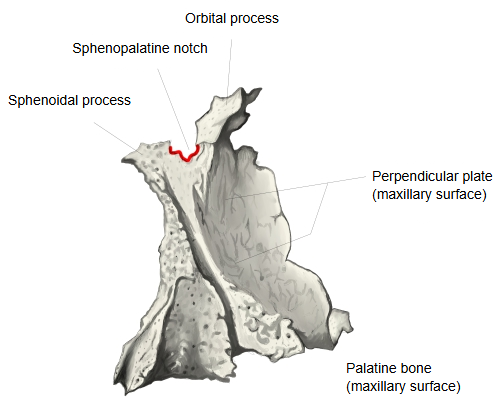
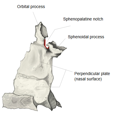
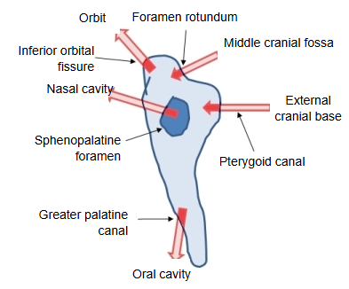
In the nasal cavity the foramen opens at the back of the superior nasal meatus, just behind the middle nasal concha; on the other side it opens into the medial wall of the pterygopalatine fossa.
The sphenopalatine artery is the main source of bleeding in posterior nosebleeds, and it can be tied or clipped at this foramen.
Reference websites
[Wikipedia]
· Sphenopalatine foramen, Wikipedia.
· Pterygopalatine fossa, Wikipedia.
[Websites with illustrations]
· Medial wall of the left orbit (Gray's Anatomy, fig. 192) (Wikimedia Commons)

· Left palatine bone, nasal aspect (Gray's Anatomy, fig. 168) (Wikimedia Commons)

Where it is found
Related terms
- Orbital process
- Sphenoidal process
- Perpendicular plate
- Body of sphenoid
- Nasal cavity
- Greater palatine canal
- Palatine bone
References and sources
- Further reading: Sphenopalatine foramen, Wikipedia.
- Further reading: Pterygopalatine fossa, Wikipedia.
- Federative International Programme for Anatomical Terminology (FIPAT). Terminologia Anatomica, 2nd ed. (TA2), 2019.