Cerebral surface of squamous part of temporal bone (Latin: facies cerebralis partis squamosae ossis temporalis)
Also called the internal surface of the temporal squama.
· The cerebral surface is the inner surface of the squamous part (temporal squama) of the temporal bone: the surface that faces the cerebrum (the temporal lobe). It is also called the internal surface. It lies on the opposite side of the squama from the outer temporal surface and forms part of the lateral wall of the middle cranial fossa.
· In short, it is the inner surface of the squama that faces the brain.
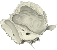 Squamous part, cerebral surface (medial view)
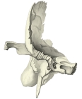 Squamous part, cerebral surface (anterior view)
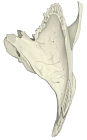 Squamous part, cerebral surface (superior view)
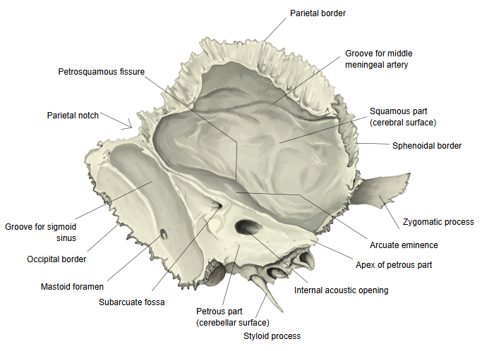
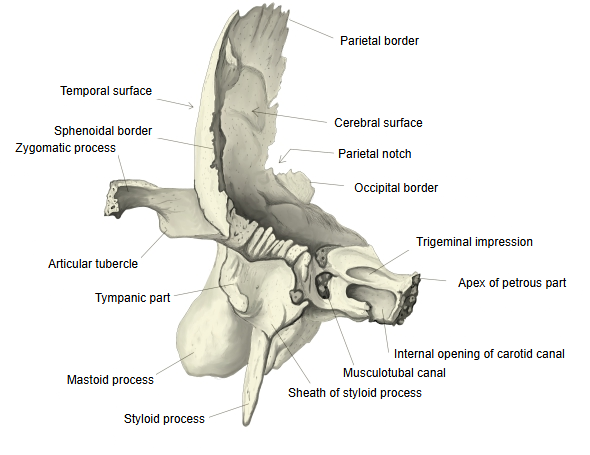
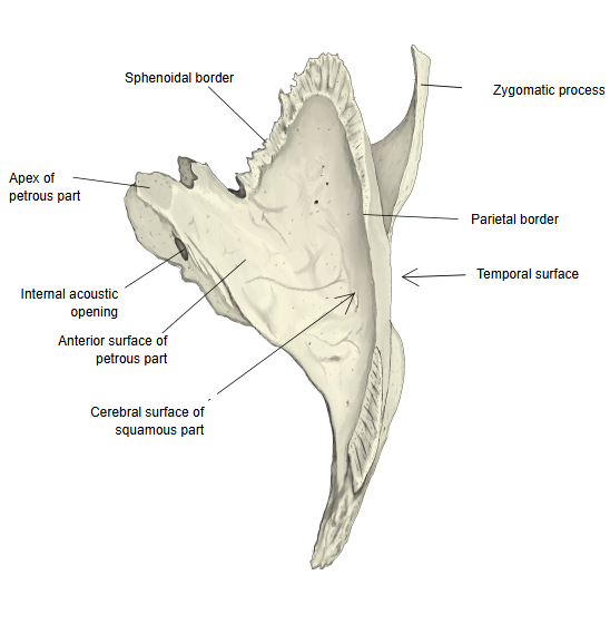
Shape and surface features
· It is slightly concave and shows digital impressions that match the gyri of the temporal lobe, with low ridges (cerebral juga) between them.
· Grooves made by the branches of the middle meningeal artery and veins run across it: arterial grooves such as the groove for the middle meningeal artery, and venous grooves.
· The groove for the middle meningeal veins is often especially clear. Between this surface and the petrous part runs the petrosquamous fissure, the trace of the fusion of the petrous and squamous parts, which becomes less distinct with age.
Boundaries
· Below, it is separated from the petrous part by the petrosquamous fissure. The fissure is clear in young people and gradually fades with age.
· Its upper (parietal) border is thin and bevelled on its inner side; it overlaps the lower border of the parietal bone to form the squamous suture. In front, the sphenoidal border joins the greater wing of the sphenoid.
· The anterior surface of the petrous part is sometimes also called a cerebral surface, but it is a different surface from the cerebral surface of the squamous part.
Clinical significance
· The squamous part is thin, so a hard blow to the temporal region can fracture it and tear the middle meningeal artery running on its cerebral surface. Blood then collects between the bone and the dura mater (extradural or epidural haematoma).
Further reading
· See the pages squamous part (temporal bone), middle cranial fossa and petrosquamous fissure.
· For the cerebral surfaces of other bones, see cerebral surface of frontal bone and cerebral surface of parietal bone.
Reference websites
[Wikipedia]
· Squamous part of temporal bone, Wikipedia.
· Temporal bone, Wikipedia.
[Websites with illustrations]
· Left temporal bone, inner surface (Gray's Anatomy, fig. 138) (Wikimedia Commons)

· Base of the skull, inner surface (Gray's Anatomy, fig. 193) (Wikimedia Commons)

Related terms
- Squamous part
- Temporal bone
- Middle cranial fossa
- Petrosquamous fissure
- Squamous suture
- Digital impressions
- Arterial grooves
- Cerebral surface of parietal bone
- Cerebral surface of frontal bone
References and sources
- Further reading: Squamous part of temporal bone, Wikipedia.
- Federative International Programme for Anatomical Terminology (FIPAT). Terminologia Anatomica, 2nd ed. (TA2), 2019.