Squamous part (Latin: pars squamosa ossis temporalis)
Also called the temporal squama or squamous part of the temporal bone
· As its name says (Latin "squama", a scale), it is thin and scale-like; much of it is thin enough to be translucent when held up to the light.
- Squamous part : occupies the middle and the whole upper part of the bone; it has two surfaces, temporal and cerebral.Related terms: groove for middle temporal artery, zygomatic process (articular tubercle), suprameatal spine
- Petrous part : the lower part, made of two parts: the mastoid part (from the mastoid process up to the parietal notch) and the pyramid (part of the cranial base).
- Tympanic part : the smallest part: a thin, half-tube-shaped plate that forms the front and lower walls of the external acoustic meatus.
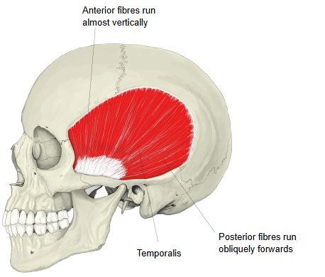
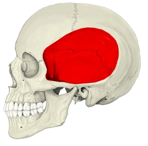
| 1 | Outer surface | = temporal surface • Smooth and convex. • Gives origin to temporalis. • Forms part of the temporal fossa. • A vertical groove for the middle temporal artery runs up its back part. • A curved ridge, the supramastoid crest, continues backwards from the posterior root of the zygomatic process; it gives attachment to the temporal fascia and marks the lower limit of temporalis. • The zygomatic process projects forwards, almost horizontally, from just above the external acoustic opening and joins the temporal process of the zygomatic bone to form the zygomatic arch. Masseter arises from its lower border and medial surface. • A small pointed projection at the upper edge of the entrance of the external acoustic meatus, when clearly present, is the suprameatal spine. |
| 2 | Inner surface | = cerebral surface Slightly concave, with ridges and impressions made by the gyri of the brain and grooves for arteries and veins; the grooves for the branches of the middle meningeal vessels are especially clear. It forms part of the floor and side wall of the middle cranial fossa. Between the cerebral surface and the petrous part runs the petrosquamous fissure, the line of fusion of the two parts; it becomes less distinct with age. |
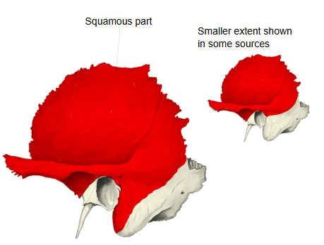
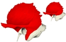 Squamous part (lateral view)
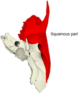
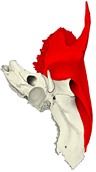 Squamous part (inferior view)
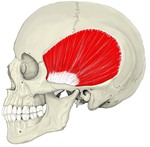 Temporalis
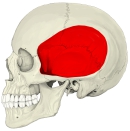 Temporal fossa
Its two borders are as follows.
| 1 | Superior border | • Thin, and bevelled on its inner side. • It overlaps the lower (squamosal) border of the parietal bone ⇒ squamous suture. • At its back end it forms the parietal notch with the mastoid part; the mastoid angle of the parietal bone fits into it. | 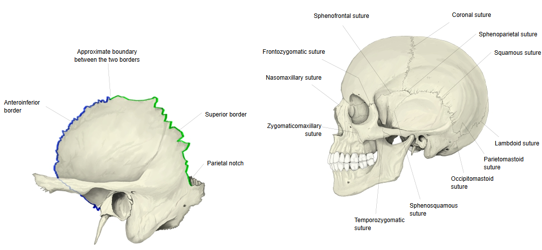 |
| 2 | Anteroinferior border | • Thick and serrated. • It joins the greater wing of the sphenoid bone ⇒ sphenosquamous suture. |
Reference websites
[Wikipedia]
· Squamous part of temporal bone, Wikipedia.
· Temporal bone, Wikipedia.
[Websites with illustrations]
· The temporal bone (TeachMeAnatomy)
· Temporal bone: anatomy, parts, sutures and foramina (Kenhub)
· Left temporal bone, outer surface (Gray's Anatomy, fig. 137) (Wikimedia Commons)

· Left temporal bone, inner surface (Gray's Anatomy, fig. 138) (Wikimedia Commons)

Surfaces
Temporal (outer) surface: smooth and convex; it forms part of the temporal fossa and gives origin to temporalis. On its back part runs a vertical groove for the middle temporal artery. A curved ridge, the supramastoid crest, runs backwards and upwards across its back part; it gives attachment to the temporal fascia and limits the origin of temporalis. The original boundary between the squamous and mastoid parts lies about 1 cm below this ridge. The zygomatic process is joined to the squama by an anterior root, which ends in the articular tubercle, and a posterior root, which passes above the external acoustic meatus into the supramastoid crest; its upper border gives attachment to the temporal fascia. The mandibular fossa of the temporomandibular joint lies on the under side of the squama. Cerebral (inner) surface: concave, with impressions of the gyri and grooves for branches of the middle meningeal vessels.
Borders
Superior border: thin and bevelled at the expense of the inner table, so that it overlaps the squamosal border of the parietal bone, forming the squamous suture. At the back it meets the mastoid part at an angle, the parietal notch. Anteroinferior border: thick and serrated, bevelled on the inner side above and on the outer side below, for the joint with the greater wing of the sphenoid bone (sphenosquamous suture). The squamous part is defined slightly differently in different atlases, so its extent in figures varies.
Where it is found
Related terms
References and sources
- Further reading: Squamous part of temporal bone, Wikipedia.
- Further reading: Temporal bone, Wikipedia.
- Federative International Programme for Anatomical Terminology (FIPAT). Terminologia Anatomica, 2nd ed. (TA2), 2019.