Corniculate tubercle (Latin: tuberculum corniculatum)
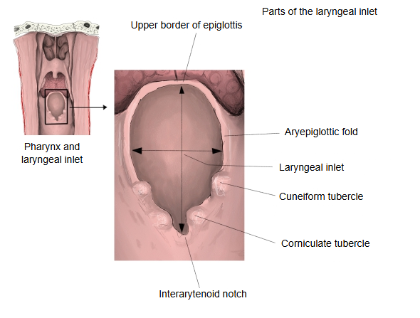
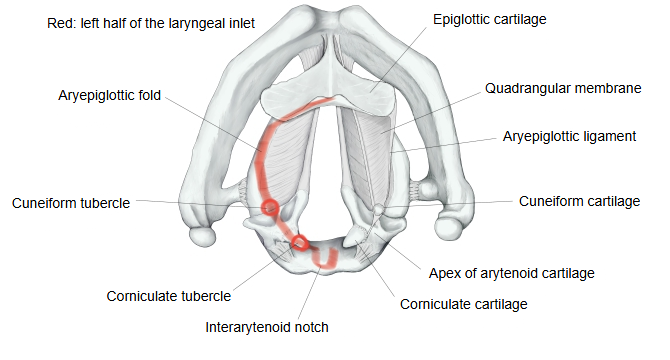
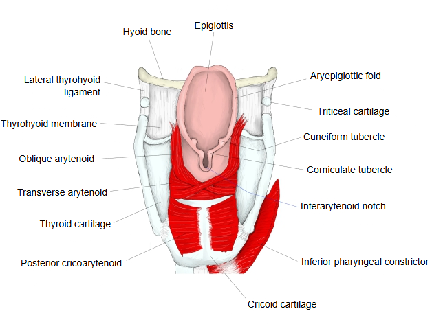
· The corniculate cartilage is a tiny cone of elastic cartilage on the apex of the arytenoid cartilage, so the tubercle marks the top of the arytenoid. It lies behind and medial to the cuneiform tubercle, just beside the interarytenoid notch that separates the right and left tubercles.
· Together with the arytenoid apices and the interarytenoid notch, the corniculate tubercles form the back edge of the laryngeal inlet.
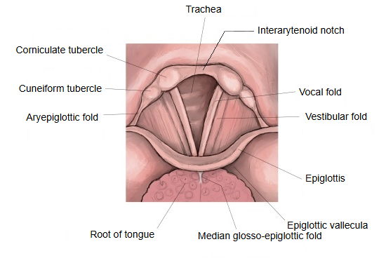
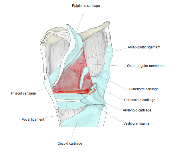
· The tubercles move with the arytenoid cartilages, so on laryngoscopy they show whether the vocal folds (glottis, rima glottidis) are opening and closing normally. The cricocorniculate ligament links the corniculate cartilage to the cricoid cartilage.
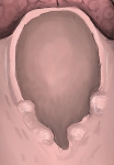 Laryngeal inlet (from behind)
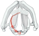 Laryngeal cartilages and the inlet
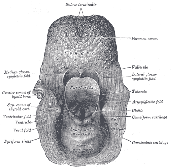
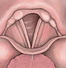 Larynx from above
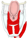 Back of the larynx
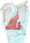 Right quadrangular membrane
Reference websites
[Wikipedia]
· Corniculate cartilages, Wikipedia.
[Websites with illustrations]
· Laryngeal cartilages (illustration) (Springer Nature)

· Entrance to the larynx seen from behind (Gray's Anatomy, fig. 955) (Wikimedia Commons)
· The larynx (TeachMeAnatomy)
Where it is found
Related terms
References and sources
- Further reading: Corniculate cartilages, Wikipedia.
- Federative International Programme for Anatomical Terminology (FIPAT). Terminologia Anatomica, 2nd ed. (TA2), 2019.