Cuneiform tubercle (Latin: tuberculum cuneiforme)
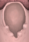 Laryngeal inlet (from behind)
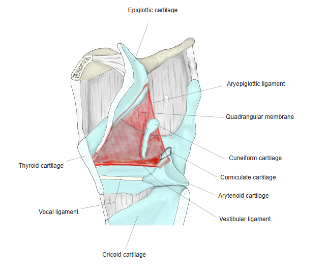
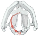 Laryngeal cartilages and the inlet
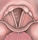 Larynx from above
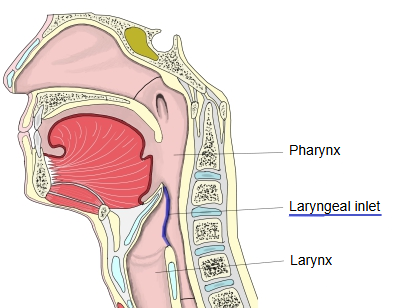
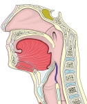 Pharynx and larynx (median section)
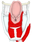 Muscles of the larynx (from behind)
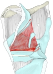 Right quadrangular membrane
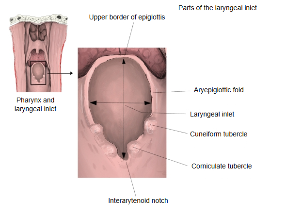
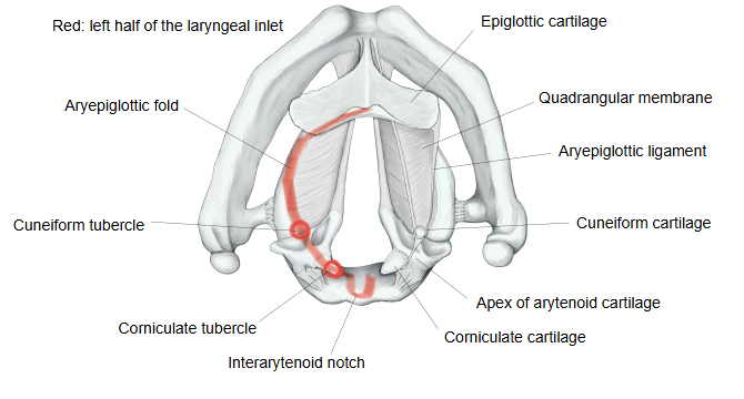
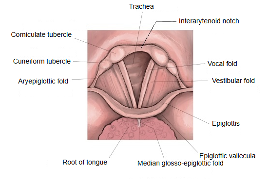
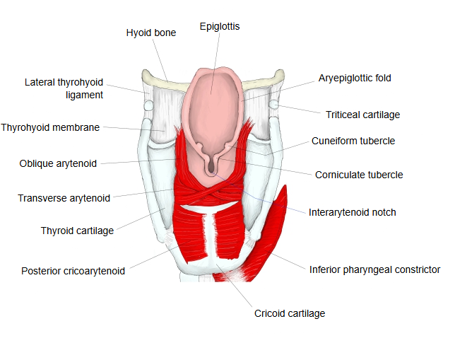
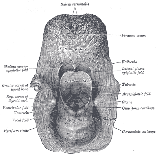
It is the larger and more lateral of the two rounded, whitish swellings on the back part of the aryepiglottic fold, raised by the cuneiform cartilage beneath it.
· The cuneiform cartilage is a small rod of elastic cartilage that is not joined to the other laryngeal cartilages. It lies in the fold beside the quadrangular membrane, stiffens the fold and helps keep the laryngeal inlet open. Its size varies, and it may be absent.
· The two tubercles help form the side and back edges of the laryngeal inlet.
· On laryngoscopy, the cuneiform and corniculate tubercles are seen as small bumps behind the vocal folds and are used as landmarks for the back of the glottis, for example during intubation.
Reference websites
[Wikipedia]
· Cuneiform cartilages, Wikipedia.
[Websites with illustrations]
· Laryngeal cartilages (illustration) (Springer Nature)

· Entrance to the larynx seen from behind (Gray's Anatomy, fig. 955) (Wikimedia Commons)
· The larynx (TeachMeAnatomy)
Where it is found
Related terms
References and sources
- Further reading: Cuneiform cartilages, Wikipedia.
- Federative International Programme for Anatomical Terminology (FIPAT). Terminologia Anatomica, 2nd ed. (TA2), 2019.