Epicranial aponeurosis (Latin: aponeurosis epicranialis; galea aponeurotica)
Also called: galea aponeurotica
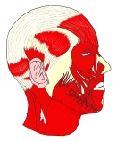 Epicranial aponeurosis (lateral view)
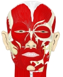 Epicranial aponeurosis (anterior view)
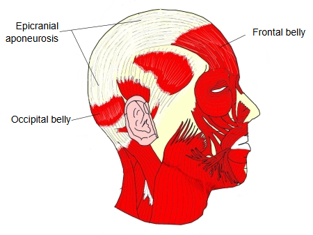
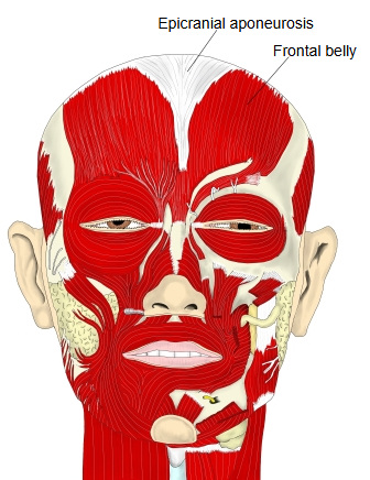
The thin, broad tendinous sheet that joins the frontalis and the occipitalis, the frontal and occipital bellies of the occipitofrontalis, part of the epicranius. It covers the top of the skull over the frontal, parietal and occipital bones; at the sides it gives attachment to the temporoparietalis, which with the occipitofrontalis makes up the epicranius, the muscle layer of the scalp.
The epicranius and the epicranial aponeurosis are firmly bound to the skin of the scalp above by dense connective tissue, but only loosely joined to the periosteum of the skull (pericranium) below, through a layer of loose areolar tissue. Because of this loose layer, the skin and the aponeurosis move together over the surface of the skull when the frontal and occipital bellies contract. It is also the plane in which the scalp separates in scalping injuries and in which blood or infection can spread across the skull.
Reference websites
[Wikipedia]
· Epicranial aponeurosis, Wikipedia.
· Scalp, Wikipedia.
[Websites with illustrations]
· Front and side views of the muscles of facial expression, with the epicranial aponeurosis (OpenStax) (Wikimedia Commons)

· Muscles of the head, face and neck (Gray's Anatomy) (Wikimedia Commons)

Related terms
References and sources
- Further reading: Epicranial aponeurosis, Wikipedia.
- Further reading: Scalp, Wikipedia.
- Federative International Programme for Anatomical Terminology (FIPAT). Terminologia Anatomica, 2nd ed. (TA2), 2019.