Epiglottic cartilage (Latin: cartilago epiglottica)
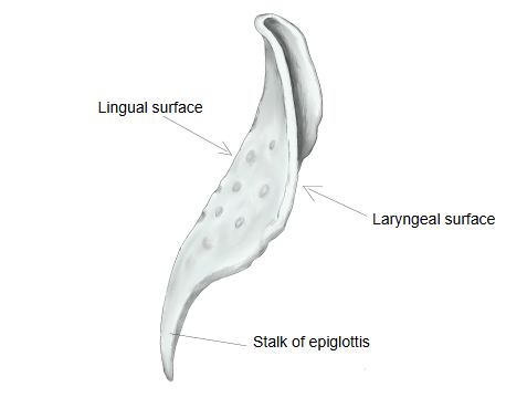
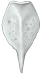 Epiglottic cartilage
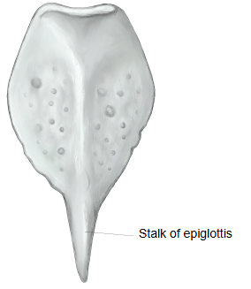
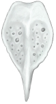 Epiglottic cartilage (posterior view)
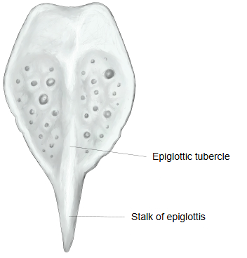
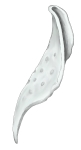 Epiglottic cartilage (lateral view)
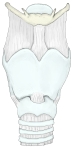 Larynx (anterior)
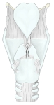 Larynx (posterior)
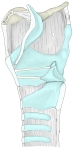 Larynx (side view, partly opened)
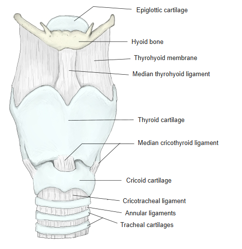
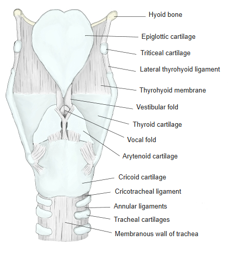
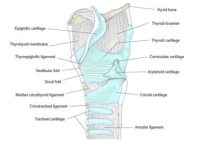
Below is a simple list of the laryngeal cartilages.
- Unpaired
- Thyroid cartilage : the largest of the laryngeal cartilages; it forms most of the front wall of the larynx (the laryngeal prominence). Hyaline cartilage.
- Cricoid cartilage : the lowest of the laryngeal cartilages; joined to the trachea by the cricotracheal ligament. Hyaline cartilage.
- Epiglottic cartilage : forms the framework of the epiglottis. Elastic cartilage.
- Paired
- Arytenoid cartilage : sits on the upper border of the cricoid lamina; the most important cartilage for voice production. Mostly hyaline cartilage.
- Corniculate cartilage : a very small cartilage on the apex of the arytenoid cartilage. Elastic cartilage.
- Cuneiform cartilage : a small flat cartilage in front of the corniculate cartilage; it may be absent. Elastic cartilage. (Some sources do not count these last two among the laryngeal cartilages.)
- Sesamoid cartilages : small, inconstant cartilages; sometimes several are present.
A standard anatomy textbook describes the epiglottic cartilage as follows.
It is unpaired and made of elastic cartilage. It is the flat cartilage that forms the base of the epiglottis, shaped like a spoon with a hollowed back. It lies at the upper opening of the larynx, against the root of the tongue. Its lower part narrows like a stalk into the stalk of the epiglottis, which is joined by the thyroepiglottic ligament to the midline of the back of the thyroid cartilage laminae. Above the stalk, a small elevation of the mucosa on the back surface is the epiglottic tubercle. The front surface is convex and faces the tongue, so it is called the lingual surface; the back surface is concave and faces the larynx, so it is called the laryngeal surface.
Reference websites
[Wikipedia]
· Epiglottis, Wikipedia.
· Larynx, Wikipedia.
[Websites with illustrations]
· Epiglottic cartilage: anterior and posterior surfaces (blogspot.com)

· Cartilages and ligaments of the larynx, posterior view (Latin labels) (en.shram.kiev.ua)

· Larynx in median section (corpshumain.ca)

· The cartilages of the larynx, posterior view (Gray's Anatomy) (Wikimedia Commons)

Structure
The epiglottic cartilage is one of the three unpaired cartilages of the larynx, with the thyroid cartilage and the cricoid cartilage. It lies at the entrance of the larynx, behind the root of the tongue and the hyoid bone. Its broad upper part is free; its lower end narrows into the stalk, which is attached by the thyroepiglottic ligament to the inner surface of the thyroid cartilage, just below the superior thyroid notch. The hyoepiglottic ligament joins its front surface to the body of the hyoid bone, and the aryepiglottic folds run from its sides back to the arytenoid cartilages. The mucosa-covered upper part of the lingual surface forms the front wall of the epiglottic valleculae.
Microscopic structure: the plate is elastic cartilage, so it stays flexible and does not ossify with age, unlike the thyroid and cricoid cartilages. It is pitted with small holes that hold mucous glands. The lingual surface is covered by non-keratinized stratified squamous epithelium, like the back of the tongue; the lower laryngeal surface by ciliated columnar (respiratory) epithelium with goblet cells.
Development: the epiglottis develops from the fourth pharyngeal arch and becomes recognizable later than the other laryngeal cartilages. In infants it lies higher and touches the soft palate; in adults it lies lower.
Function
During breathing the epiglottis points upward and air passes freely into the larynx. Its mucosa carries some taste buds.
Swallowing: when the hyoid bone and the larynx rise and move forward during swallowing, the epiglottis bends back over the laryngeal inlet, helping to keep food out of the trachea and steer it into the oesophagus behind. Pressure from the tongue, the weight of the food and the small muscles of the laryngeal inlet all contribute to this movement. Speech: in most languages the epiglottis plays no part in forming sounds; a few languages use it for rare epiglottal consonants.
Related terms
- Epiglottis
- Thyroid cartilage
- Arytenoid cartilage
- Epiglottic vallecula
- Thyroepiglottic ligament
- Hyoepiglottic ligament
- Larynx
References and sources
- Further reading: Epiglottis, Wikipedia.
- Further reading: Larynx, Wikipedia.
- Federative International Programme for Anatomical Terminology (FIPAT). Terminologia Anatomica, 2nd ed. (TA2), 2019.