Epiglottis (Latin: epiglottis)
· The epiglottis carries taste buds, small groups of cells that sense taste.
· In the living body it looks almost white.
· It rises behind the root of the tongue and curves backward, with its free tip pointing upward and backward.
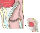 Larynx (median section)
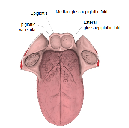
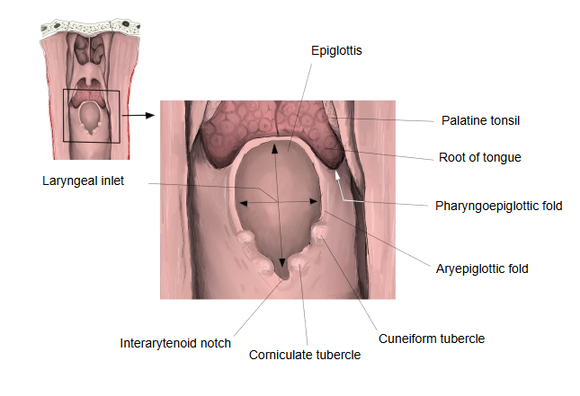
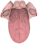 Tongue and epiglottis
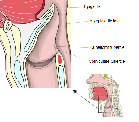
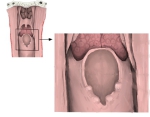 Laryngeal inlet (posterior view)
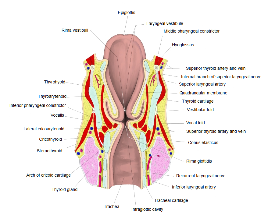
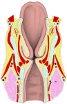 Larynx (coronal section)
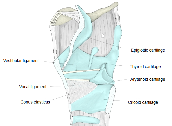
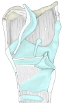 Cartilages of the larynx (median section)
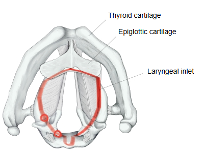
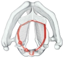 Laryngeal inlet (superior view)
Reference websites
[Wikipedia]
· Epiglottis, Wikipedia.
[Websites with illustrations]
· The entrance to the larynx, viewed from behind (Gray's Anatomy) (Wikimedia Commons)

· Sagittal section of the larynx (Gray's Anatomy) (Wikimedia Commons)

· Normal larynx: cut-away view and view from above (anatomyexhibits.com)

· Epiglottis in anterior, posterior and median views (healthfixit.com)

Structure
The epiglottis is a leaf-shaped flap at the entrance of the larynx, formed by the epiglottic cartilage, a plate of elastic cartilage covered with mucous membrane. Its narrow lower stalk is attached to the inner surface of the thyroid cartilage, just below the notch; the broad upper part rises behind the root of the tongue and the hyoid bone.
Its front surface is joined to the tongue by the median and lateral glosso-epiglottic folds; the hollows between them are the epiglottic valleculae. From its sides the aryepiglottic folds run back to the arytenoid cartilages and frame the laryngeal inlet.
Function
During breathing the epiglottis stands upright and air passes into the larynx. During swallowing the hyoid bone and the larynx rise and the epiglottis tips back over the inlet, helping to steer food and liquid behind the larynx into the oesophagus instead of the trachea.
Related terms
- Larynx
- Aryepiglottic fold
- Epiglottic vallecula
- Lateral glosso-epiglottic fold
- Pharyngo-epiglottic fold
- Laryngeal vestibule
- Hyoid bone
References and sources
- Further reading: Epiglottis, Wikipedia.
- Federative International Programme for Anatomical Terminology (FIPAT). Terminologia Anatomica, 2nd ed. (TA2), 2019.