Foramen lacerum (Latin: foramen lacerum)
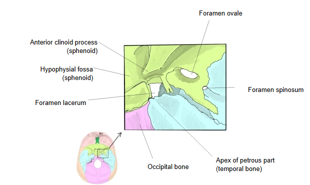
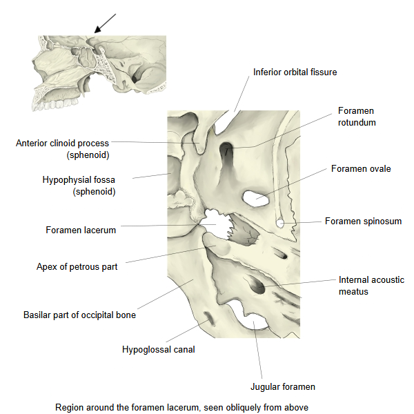
· It is an irregular gap between the body of the sphenoid and the petrous part (apex) of the temporal bone.
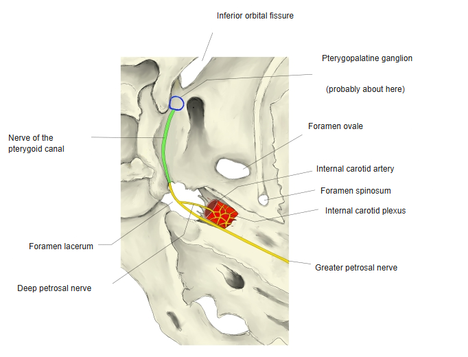
It lies at the medial end of the petrous part, just in front of and medial to the internal opening of the carotid canal, where the sphenopetrosal and petro-occipital fissures meet (the name means 'torn opening'). In life its lower part is closed by a plate of fibrocartilage. The internal carotid artery leaves the carotid canal and runs across the upper surface of the cartilage, then turns up into the cavernous sinus. The anterior margin of the foramen leads into the pterygoid canal: on the cartilage the greater petrosal nerve (parasympathetic) and the deep petrosal nerve (sympathetic, from the internal carotid plexus) join to form the nerve of the pterygoid canal, which runs forward into the canal with the artery of the pterygoid canal. Small emissary veins may link the cavernous sinus with the pterygoid venous plexus through the cartilage.
This description can be read in two ways.
1. 1. The foramen itself is closed by cartilage and nothing passes through it ⇒ no nerves or vessels pass through the foramen lacerum.
2. 2. Between part of its anterior margin and the sphenoid there is a gap, and nerves and vessels pass through it
⇒ (not in the strict sense) nerves and vessels do pass through the foramen lacerum
Sources that say they pierce the cartilage of the foramen lacerum:
· The greater petrosal nerve pierces the cartilage of the foramen lacerum to reach the cranial base.
· The greater petrosal nerve pierces the cartilage of the foramen lacerum and comes out on the outer surface of the cranial base.
· In life the foramen lacerum is closed by cartilage. Part of its anterior margin is formed by the sphenoid, and there the internal carotid artery turns upwards into the cavernous sinus.
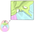 Bones around the foramen lacerum (colour-coded)
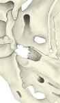 Foramen lacerum region (oblique view from above)
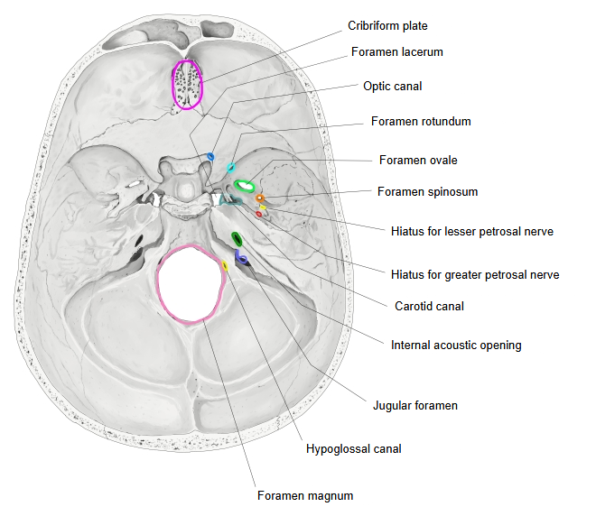
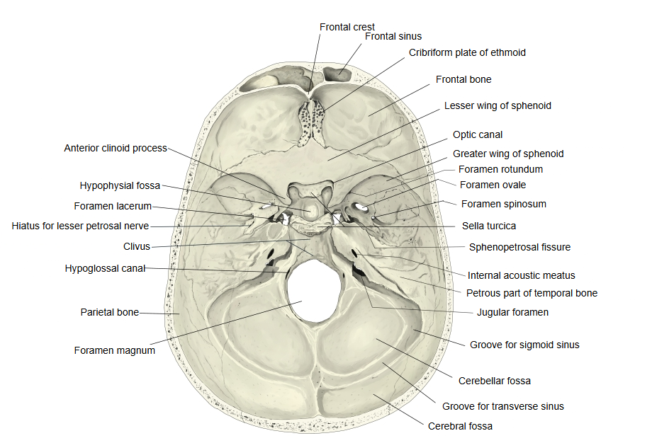
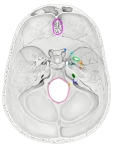 Openings of the cranial base
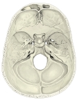 Cranial base (internal surface)
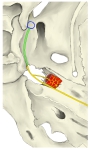 Course of the nerve of the pterygoid canal
[Vessels and nerves passing through it]
Textbooks differ here: some descriptions of the foramen lacerum mention no vessels or nerves passing through it, while others (for example the Prometheus atlas) list the greater petrosal nerve and the deep petrosal nerve.
| 1 | Nerves | Greater petrosal nerve, deep petrosal nerve |
| 2 | Arteries | Artery of the pterygoid canal, posterior meningeal artery (a meningeal branch of the ascending pharyngeal artery); the latter may also pass through other openings |
| 3 | Veins | Emissary vein of the foramen lacerum (one of the emissary veins) |
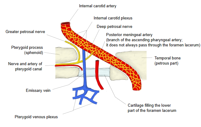
About the emissary vein of the foramen lacerum in the table above, textbooks say:
Through the foramen lacerum it connects the cavernous sinus with the pterygoid venous plexus.
Reference websites
[Wikipedia]
· Foramen lacerum, Wikipedia.
· Artery of pterygoid canal, Wikipedia.
[Websites with illustrations]
· Base of the skull, inner surface (Gray's Anatomy, fig. 193) (Wikimedia Commons)

· Foramen lacerum (Kenhub)
Where it is found
Related terms
- Carotid canal
- Petrous part
- Internal carotid artery
- Greater petrosal nerve
- Deep petrosal nerve
- Nerve of pterygoid canal
- Emissary vein of foramen lacerum
References and sources
- Further reading: Foramen lacerum, Wikipedia.
- Federative International Programme for Anatomical Terminology (FIPAT). Terminologia Anatomica, 2nd ed. (TA2), 2019.