Iliopectineal arch (Latin: arcus iliopectineus)
What is the iliopectineal arch?
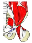 Muscular and vascular lacunae (anterior view)
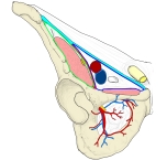 Contents of the lacunae
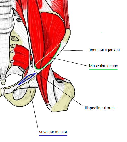
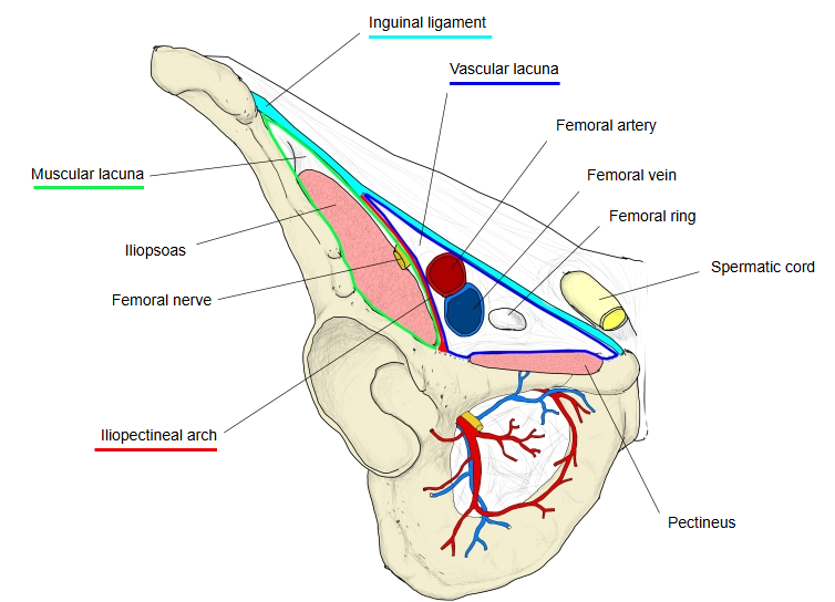
The iliopectineal arch is a thick band formed where the fasciae of the iliacus and the psoas major (the iliac fascia and the psoas fascia) fuse. In front it arises from the back of the inguinal ligament; it passes backwards between the femoral nerve (lateral to it) and the femoral vessels (medial to it) and ends behind on the iliopubic eminence of the hip bone. In this way it divides the space below the inguinal ligament into the lateral muscular lacuna and the medial vascular lacuna. When a psoas minor is present, its tendon blends with the arch.
· Lateral to it (muscular lacuna) pass the iliopsoas and the femoral nerve; medial to it (vascular lacuna) pass the femoral artery and vein.
· It is sometimes cut to release the femoral nerve when it is compressed.
Reference websites
[Wikipedia]
· Iliopectineal arch, Wikipedia.
Related terms
- Pecten pubis
- Vascular lacuna
- Inguinal ligament
- Iliopsoas
- Femoral nerve
- Psoas minor
- Hip bone
- Lacunar ligament
References and sources
- Further reading: Iliopectineal arch, Wikipedia.
- Federative International Programme for Anatomical Terminology (FIPAT). Terminologia Anatomica, 2nd ed. (TA2), 2019.