Intercondylar fossa (Latin: fossa intercondylaris)
Also called the intercondylar notch
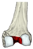 Lower right femur (posterior)
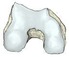 Right femur (inferior)
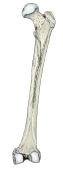 Right femur (posterior)
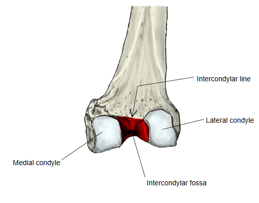
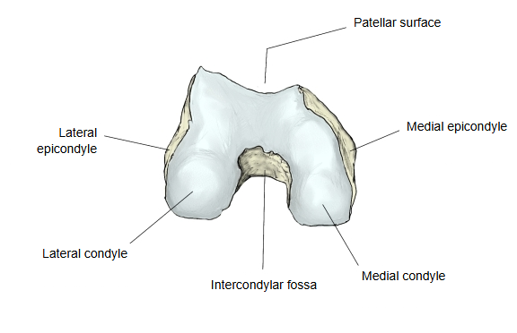
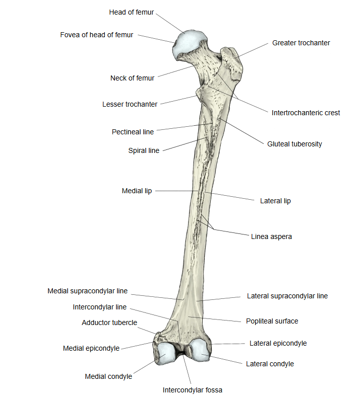
· It opens backwards and downwards; in front it is closed off by the patellar surface.
· Above, it is separated from the popliteal surface by a low ridge, the intercondylar line.
· A narrow notch, seen on imaging, is often discussed as a risk factor for anterior cruciate ligament injury.
In more detail:
It is the deep notch between the two condyles. Its lateral wall (the inner surface of the lateral condyle) has a smooth, shallow area where the anterior cruciate ligament is attached; its medial wall (the inner surface of the medial condyle) carries the attachment of the posterior cruciate ligament. Like most ligament and tendon attachment areas, these surfaces have few nutrient foramina.
 Walls and roof of the fossa
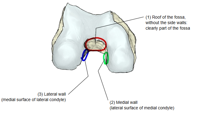
In the figure above, the red outline (1) marks the roof of the fossa, and the green (2) and blue (3) marks show its medial wall (the lateral surface of the medial condyle) and its lateral wall (the medial surface of the lateral condyle).
Does the fossa mean only the roof (1), or does it include the side walls (2 and 3)? In most English-language descriptions both side walls are counted as part of the fossa, so the cruciate ligaments are said to attach within the fossa. Sources differ on how far its boundaries extend.
Attached structures
Taking the fossa as parts 1 to 3 in the figure above:
| Ligaments | 1 | Anterior cruciate ligament (lateral wall) | ligamentum cruciatum anterius |
| 2 | Posterior cruciate ligament (medial wall) | ligamentum cruciatum posterius |
Reference websites
[Wikipedia]
· Intercondylar fossa of femur, Wikipedia.
· Femur, Wikipedia.
[Websites with illustrations]
· Lower end of the right femur from below (Gray's Anatomy, fig. 246) (Wikimedia Commons)

· Right femur, posterior surface (Gray's Anatomy, fig. 245) (Wikimedia Commons)

Where it is found
Related terms
- Intercondylar line
- Lateral condyle of femur
- Medial condyle of femur
- Popliteal surface
- Patellar surface of femur
- Distal epiphysis of femur
- Knee joint
References and sources
- Further reading: Intercondylar fossa of femur, Wikipedia.
- Further reading: Femur, Wikipedia.
- Federative International Programme for Anatomical Terminology (FIPAT). Terminologia Anatomica, 2nd ed. (TA2), 2019.