Medial condyle of femur (Latin: condylus medialis femoris)
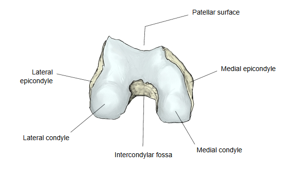
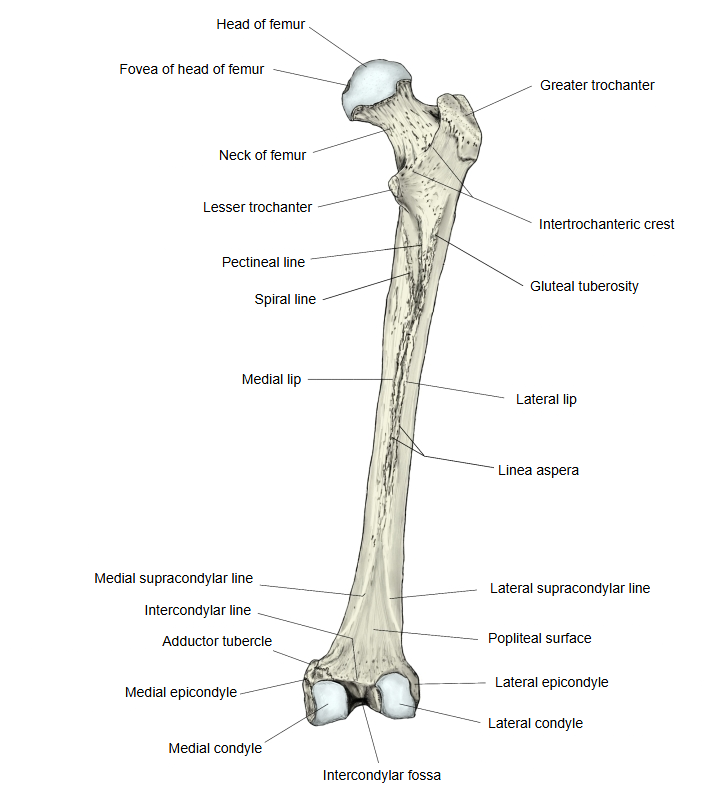
Textbooks do not define its limits precisely, but it can be understood as follows: the medial condyle is the large medial mass, the medial epicondyle is the part of it that projects most medially, and the adductor tubercle is the small projection above the epicondyle.
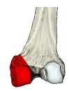 Lower right femur (posterior)
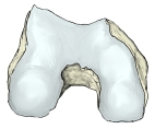 Right femur (inferior)
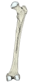 Right femur (posterior)
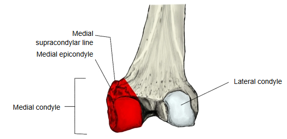
Attachments
The list below includes structures attached to the medial epicondyle and the adductor tubercle, so it overlaps with those pages.
| Muscle | 1 | Gastrocnemius (medial head, origin) | above the medial condyle, behind the medial epicondyle | musculus gastrocnemius, caput mediale |
| 2 | Adductor magnus (insertion) | adductor tubercle | musculus adductor magnus | |
| Ligament | 1 | Tibial collateral ligament | medial epicondyle | ligamentum collaterale tibiale |
| 2 | Posterior cruciate ligament | medial condyle (the surface facing the intercondylar fossa) | ligamentum cruciatum posterius |
The lower part of the shaft of the femur becomes especially thick and wide, and its lower end is formed by the two enlarged condyles, medial and lateral. Compared with the lateral condyle, the medial condyle is narrower and longer, with a more strongly curved surface, and it reaches further down.
The two condyles make up the lower end of the femur. In front they are joined by the smooth patellar surface, which articulates with the back of the patella. Behind, they are separated by a deep notch, the intercondylar fossa, which is marked off above from the popliteal surface by the intercondylar line. The lower and posterior surfaces of the medial condyle articulate with the medial condyle of the tibia, with the medial meniscus in between, at the knee joint.
The medial condyle is one of the two projections at the lower end of the femur, the other being the lateral condyle. It is often described as the larger of the two, because the line of body weight passes medial to the knee. On the back of the bone, the medial lip of the linea aspera continues down as the medial supracondylar line. The most prominent point on the medial surface of the medial condyle is the medial epicondyle, which can be felt by running the fingers medially from the patella with the knee bent.
· The medial condyle is longer and reaches further down than the lateral condyle; sources differ on which of the two is called the larger.
· On the back, the medial lip of the linea aspera continues as the medial supracondylar line, which ends at the adductor tubercle.
· The most medial projecting part is called the medial epicondyle.
Reference websites
[Wikipedia]
· Medial condyle of femur, Wikipedia.
· Femur, Wikipedia.
[Websites with illustrations]
· Lower end of the femur, posterior view (Wikimedia Commons)

· Lower end of the right femur viewed from below (Gray's Anatomy, fig. 246) (Wikimedia Commons)

· The femur (TeachMeAnatomy)
Where it is found
Related terms
- Lateral condyle of femur
- Medial epicondyle of femur
- Adductor tubercle
- Intercondylar fossa
- Patellar surface of femur
- Distal epiphysis of femur
- Knee joint
References and sources
- Further reading: Medial condyle of femur, Wikipedia.
- Further reading: Knee, Wikipedia.
- Federative International Programme for Anatomical Terminology (FIPAT). Terminologia Anatomica, 2nd ed. (TA2), 2019.