Distal epiphysis of femur (Latin: epiphysis distalis femoris)
Also called the lower end of the femur
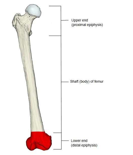
The distal epiphysis is one of the three parts into which the femur is divided: the greatly enlarged lower end of the bone. It is also called the lower end of the femur.
- Proximal epiphysis : marked by the projecting head and neck of the femur. = upper end
- Shaft (body) of femur : the tubular part that forms the shaft of the bone.
- Distal epiphysis : much more enlarged than the proximal end. = lower end
The lower end of the femur is broadly swollen. The inner swelling is the medial condyle and the outer one the lateral condyle. The medial condyle bulges downwards, inwards and backwards; the bump on its inner side is the medial epicondyle, with the small adductor tubercle just above it. The lateral condyle bulges less to the side and hardly projects beyond the outer surface of the shaft; the bump on its outer side is the lateral epicondyle. In front the two condyles are joined, but behind they are separated by a deep gap, the intercondylar fossa, whose upper edge is the ridge called the intercondylar line. The convex lower and posterior surfaces of the condyles form the articular surface for the upper end of the tibia; the medial part curves forwards and outwards, while the lateral part is broad and runs straight from front to back. In front of the condyles the two parts merge into the patellar surface, which meets the articular surface on the back of the patella. A vertical groove divides the patellar surface into medial and lateral parts, the lateral part being larger.
· The lower and posterior surfaces of the condyles articulate with the tibia, with the menisci in between, forming the upper part of the knee joint.
The distal epiphysis is made up of the following parts.
| 1 | Lateral condyle | condylus lateralis | 6 | Medial condyle | condylus medialis |
| 2 | Lateral supracondylar line | linea supracondylaris lateralis | 7 | Medial supracondylar line | linea supracondylaris medialis |
| 3 | Lateral epicondyle | epicondylus lateralis | 8 | Medial epicondyle | epicondylus medialis |
| 4 | Patellar surface | facies patellaris | 9 | Adductor tubercle | tuberculum adductorium |
| 5 | Groove for popliteus | sulcus popliteus |
The following muscles attach to the distal epiphysis (red: origin, blue: insertion). Some also reach the shaft of the femur.
| 1 | Popliteus (groove for popliteus, just below the lateral epicondyle) | 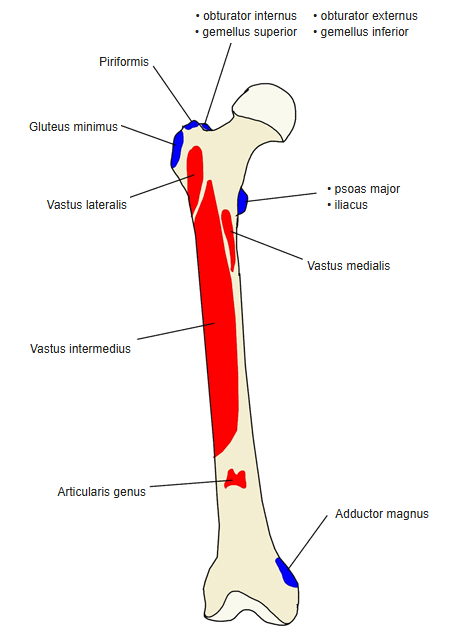 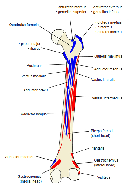 |
| 2 | Articularis genus (front of the lower shaft, just above the patellar surface) | |
| 3 | Plantaris (lower end of the lateral supracondylar line) | |
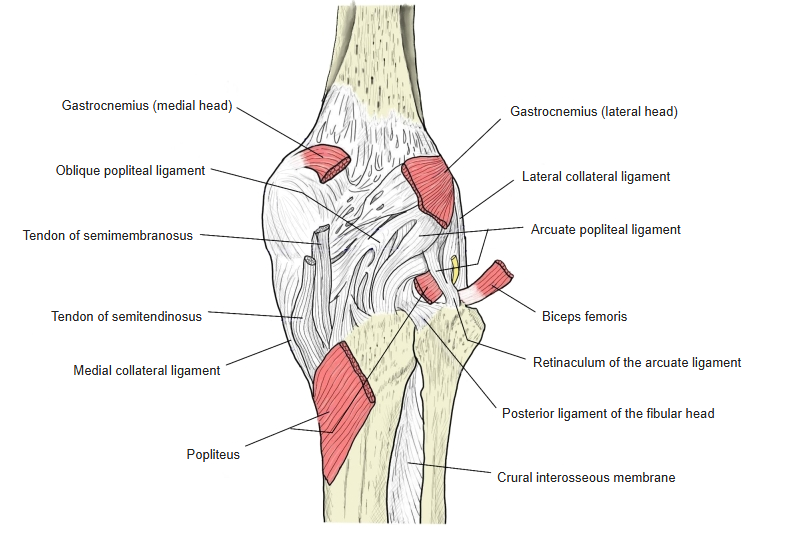
| 4 | Gastrocnemius (above the medial condyle; lateral surface of the lateral condyle) | |
| 1 | Adductor magnus (medial supracondylar line, adductor tubercle) | |
The following ligaments attach to the distal epiphysis.
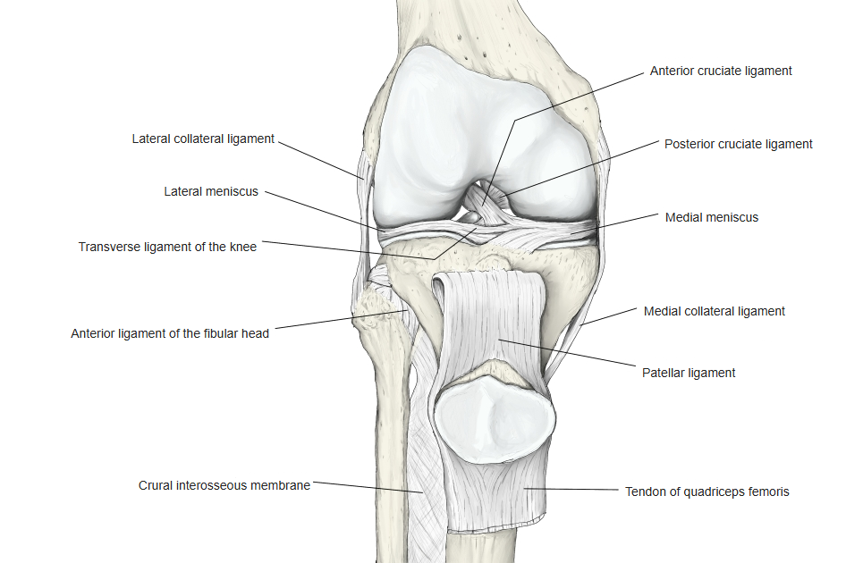
| 1 | Fibular collateral ligament | Femur (lateral epicondyle) ⇔ fibula (head of fibula) |
| 2 | Tibial collateral ligament | Femur (medial epicondyle) ⇔ tibia (proximal epiphysis: medial condyle, and the upper shaft) |
| 3 | Anterior cruciate ligament | Femur (medial surface of the lateral condyle, facing the intercondylar fossa) ⇔ tibia (proximal epiphysis: anterior intercondylar area) |
| 4 | Posterior cruciate ligament | Femur (lateral surface of the medial condyle, facing the intercondylar fossa) ⇔ tibia (proximal epiphysis: posterior intercondylar area). The anterior and posterior ligaments together are called the cruciate ligaments of the knee. |
| 5 | Oblique popliteal ligament | Tendon of semimembranosus ⇔ femur (back of the lateral condyle) |
| 6 | Arcuate popliteal ligament | Fibula (head of fibula) ⇔ back of the joint capsule, arching over popliteus (some sources add a limb to the lateral condyle of the femur) |
 Knee joint (anterior)
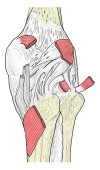 Knee joint (posterior)
Reference websites
[Wikipedia]
· Femur, Wikipedia.
· Knee, Wikipedia.
[Websites with illustrations]
· Right femur, anterior surface (Gray's Anatomy, fig. 244) (Wikimedia Commons)

· Right femur, posterior surface (Gray's Anatomy, fig. 245) (Wikimedia Commons)

· Lower end of the right femur from below (Gray's Anatomy, fig. 246) (Wikimedia Commons)

Where it is found
Related terms
- Proximal epiphysis of femur
- Shaft of femur
- Medial condyle of femur
- Lateral condyle of femur
- Patellar surface of femur
- Intercondylar fossa
- Knee joint