External acoustic meatus (Latin: meatus acusticus externus)
Also called the external auditory canal or the ear canal
- Overview
- Structure
- Curves
- Changes with age
- Vessels and nerves
- Reference websites
- (Wikipedia, websites with illustrations)
- Where it is found
- Related terms
Overview
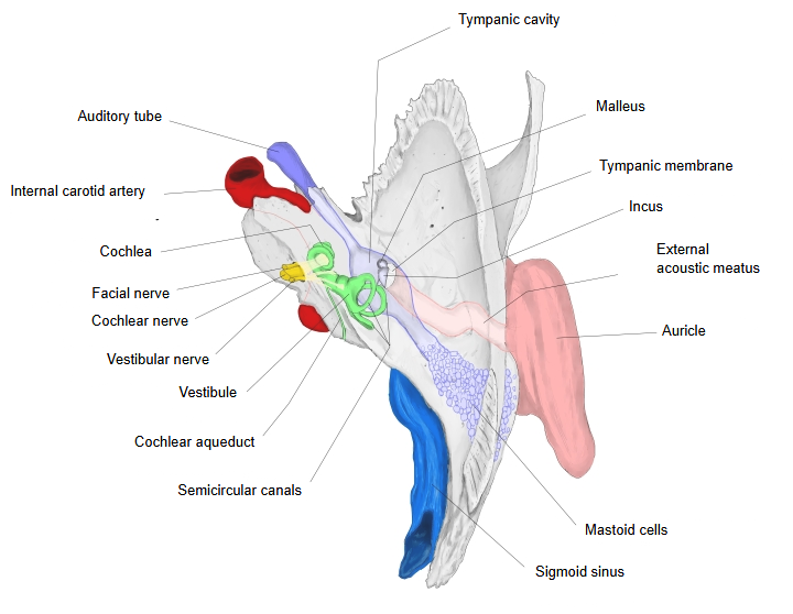
- Organ of hearing
- External ear : 1. auricle 2. external acoustic meatus 3. tympanic membrane
- Middle ear : 1. tympanic cavity 2. auditory ossicles 3. auditory tube
- Internal ear : the cochlea; its nerve leaves through the internal acoustic meatus
- Organ of balance
- Vestibule : faces the middle ear at the vestibular (oval) window; the cochlear (round) window below it opens into the cochlea
- Semicircular canals : 1. anterior 2. posterior 3. lateral semicircular canal
 Right auricle and external acoustic meatus (front view)
 Right temporal bone (superior view)
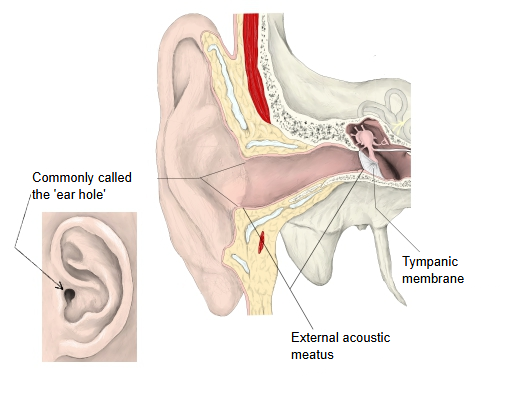
Structure
acoustic
meatus
- Cartilaginous part : the outer third of the canal (about 8 mm), made of elastic cartilage (the same as the auricular cartilage).The two parts are firmly joined by tough connective tissue.
- Bony part : the inner two-thirds of the canal (about 16 mm), in the tympanic part of the temporal bone, with the squamous part in its roof.
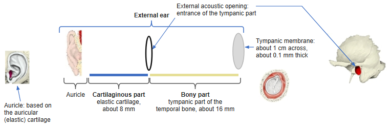
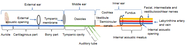
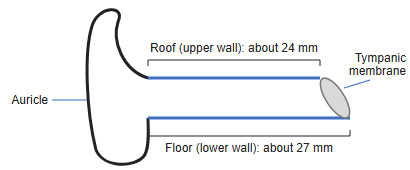
The external acoustic meatus joins the auricle to the middle ear. It is made of the outer cartilaginous part and the inner bony part: the cartilaginous part is short (about one third), the bony part long (about two thirds). The two meet at the bony external acoustic opening, and the canal is narrowest near this junction (the isthmus).
Cartilage of the meatus: a half-ring (a groove) that is open above and behind. Its inner end is attached to the rim of the external acoustic opening of the bony part, and its outer end is continuous with the cartilage of the auricle. The short gap in its back and upper wall is closed by a membrane of fibrous connective tissue.
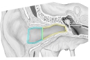 Cartilaginous and bony parts (right ear, coronal section)
- Right temporal bone (superior view)
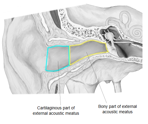
Bony part: the skin is very thin, has no glands and is firmly attached to the periosteum.
Cartilaginous part: the skin is a little thicker and contains sebaceous glands and fine hairs, and also the ceruminous glands.
Ceruminous glands: a kind of apocrine sweat gland with a fatty secretion; together with shed skin cells it forms earwax (cerumen).
At the inner end of the canal the skin becomes very thin and forms the outer (skin) layer of the tympanic membrane. The coarse hairs that grow in the canal in old age, especially in men, are called tragi. Everything above describes the adult; in children, and especially in infants, the external ear differs considerably.
Curves
The external acoustic meatus has two curves, so the tympanic membrane normally cannot be seen directly from outside. The curves lie mainly in the cartilaginous part.
First curve: just inside the entrance; here the canal runs forwards and upwards.
Second curve: near the junction of the cartilaginous and the bony part; here the canal turns backwards (and slightly upwards), and the bony part then runs forwards and slightly downwards to the tympanic membrane. Descriptions of the curves vary between sources.
Changes with age
The canal of the newborn differs from the adult one mainly in two ways: it has no bony part (the tympanic part of the temporal bone is only a ring, the tympanic ring), and the tympanic membrane is strongly tilted, lying almost horizontal. The adult canal is an open tube, but in infants the upper and lower walls nearly touch, so the canal is slit-like. To see the tympanic membrane the auricle is pulled upwards and backwards in adults, while in infants the canal opens better when the earlobe is pulled downwards (and backwards).
Vessels and nerves
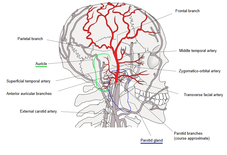
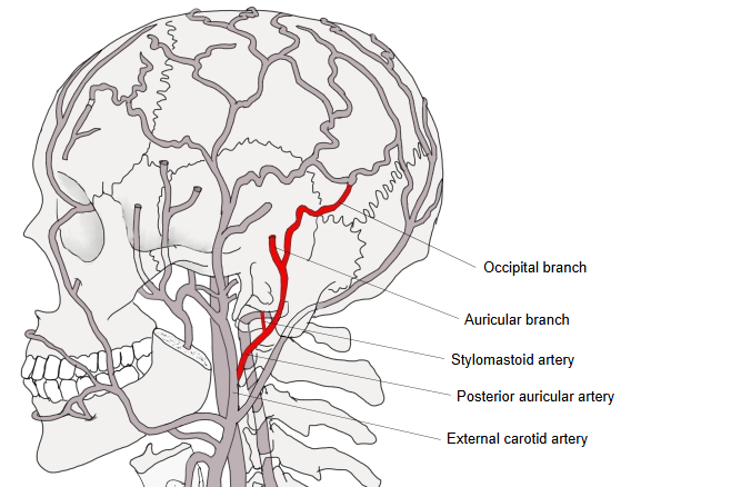
Arteries: the anterior auricular branches (from the superficial temporal artery) and the posterior auricular artery; the bony part also receives the deep auricular artery from the maxillary artery.
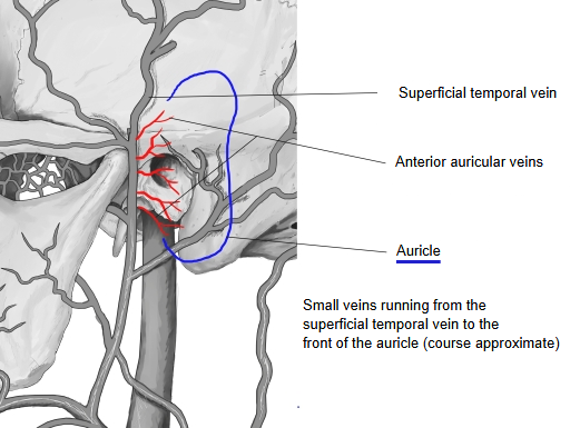
Veins: the anterior auricular veins; the blood drains to the superficial temporal, maxillary and external jugular veins.
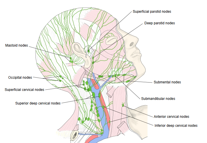
Lymph vessels: to the retroauricular (mastoid) lymph nodes, the superficial and deep parotid lymph nodes and the superior deep cervical lymph nodes.
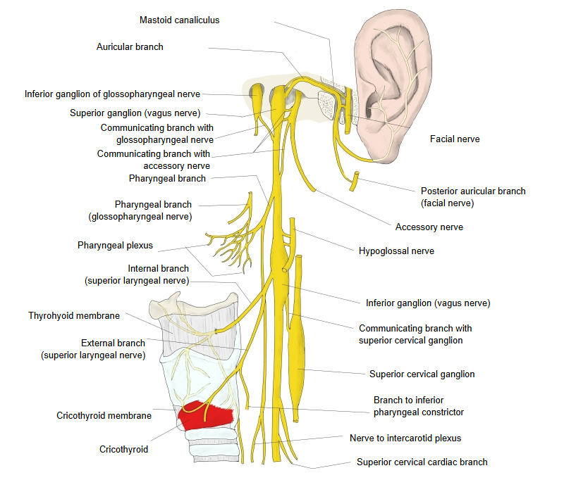
Nerves: the nerve of the external acoustic meatus from the auriculotemporal nerve (a branch of the mandibular nerve) supplies the front and roof, and the auricular branch of the vagus nerve (Arnold's nerve) supplies the back and floor. Touching the canal can therefore trigger a cough reflex.
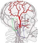 Anterior auricular branches (right temporal region)
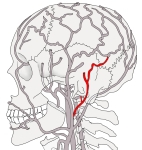 Posterior auricular artery (left temporal region)
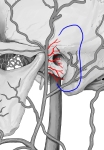 Anterior auricular veins (left temporal region)
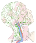 Lymph nodes of the head and neck
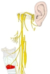 Vagus nerve (head and neck, model)
Reference websites
[Wikipedia]
· Ear canal, Wikipedia.
· Outer ear, Wikipedia.
[Websites with illustrations]
· The external ear (TeachMeAnatomy)
· Ear anatomy: parts and functions (Kenhub)
· Section through the structures of the ear (Gray's Anatomy, fig. 907) (Wikimedia Commons)
