Lacrimal canaliculus (Latin: canaliculus lacrimalis)
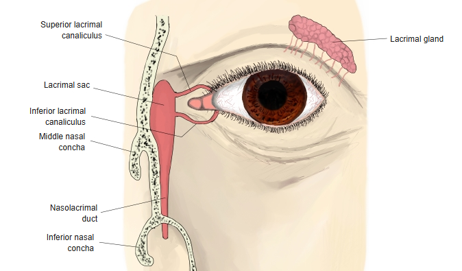
A fine duct that begins at the edge of the upper and lower eyelids near the inner corner of the eye. It is the first part of the lacrimal passage, the drainage part of the lacrimal apparatus that carries the tears to the nasal cavity.
(In the figure, the lacrimal canaliculi are shown in blue.)
More details
Each canaliculus begins at a lacrimal punctum, a tiny opening on the posterior edge of the eyelid (upper and lower) near the medial angle of the eye. It runs medially and opens into the lacrimal sac, carrying the tears to it.
There are two ways in which they open into the lacrimal sac: the upper and lower canaliculi either join first, or each opens into the sac separately.
There is one canaliculus in each eyelid, upper and lower. They bend as they run medially toward the lacrimal sac.
In most people the medial ends of the two canaliculi join to form a short common canaliculus (about 0.8–2.2 mm long), which opens into the lacrimal sac. In others the two canaliculi open separately into a small recess of the sac.
apparatus
- Lacrimal gland : secretes the tears.
- Lacrimal passage : carries the tears to the nose.
- Lacrimal canaliculus
- Lacrimal sac
- Nasolacrimal duct
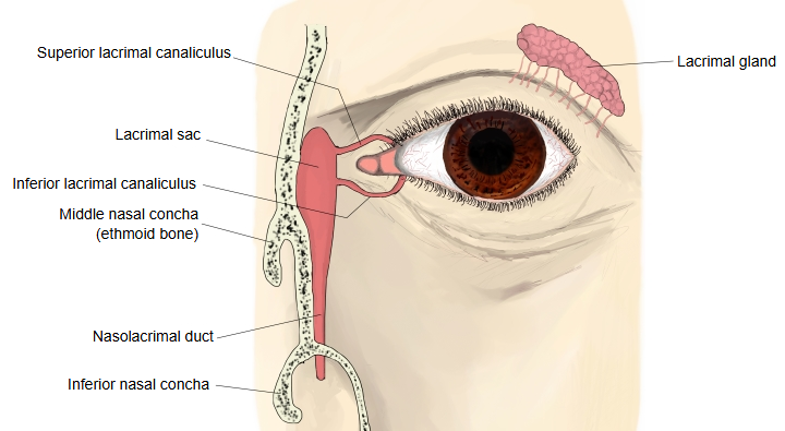
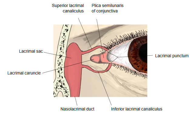
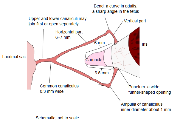
Course and width of the duct
Each canaliculus first runs vertically for about 2 mm (the upper one upward, the lower one downward) and then turns medially to run horizontally for about 6–7 mm toward the lacrimal sac. In adults the bend between the two parts is a smooth curve; in the fetus it is a sharp angle. The canaliculus begins with a wide, funnel-shaped opening at the punctum and soon narrows; beyond the narrow part it widens again at the bend to form the ampulla (inner diameter about 1 mm). The horizontal part narrows gradually and is only about 0.3 mm wide where it enters the common canaliculus (or the sac). The lower papilla (with its punctum) lies about 6.5 mm from the medial angle of the eye and the upper one about 6 mm, so the lower canaliculus is slightly longer. When the eyelids close, the lower papilla lies lateral to the upper one; both papillae point slightly backward, the upper one downward and the lower one upward.
Fine structure
The canaliculus is lined by stratified squamous epithelium, surrounded by a lamina propria of connective tissue and by longitudinal bundles of striated muscle fibres (fibres of the orbicularis oculi muscle).
The epithelium has 10–12 layers of cells and is about 120 µm thick; its deepest cells are columnar and its surface cells flat.
Reference websites
[Wikipedia]
· Lacrimal canaliculi, Wikipedia.
· Lacrimal apparatus, Wikipedia.
[Websites with illustrations]
· The lacrimal apparatus (Sobotta, 1909) (Wikimedia Commons)

· Lacrimal apparatus (illustrations and photos) (Wikimedia Commons)
Related terms
- Lacrimal caruncle
- Lacrimal passage
- Fossa for lacrimal gland
- Lacrimal groove of maxilla
- Lacrimal margin of maxilla
- Lacrimal notch
- Lacrimal process
- Lunula lacrimalis
- Lacrimal apparatus
- Lacrimal sac
- Nasolacrimal duct
- Lacrimal gland
- Edge of eyelid
- Eyelids
- Fossa for lacrimal sac
References and sources
- Further reading: Lacrimal canaliculi, Wikipedia.
- Further reading: Lacrimal apparatus, Wikipedia.
- Federative International Programme for Anatomical Terminology (FIPAT). Terminologia Anatomica, 2nd ed. (TA2), 2019.