Medial meniscus (Latin: meniscus medialis)
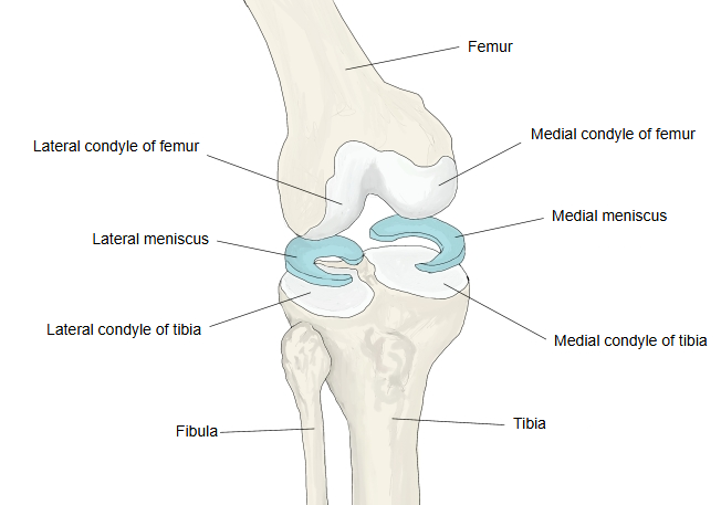
The articular meniscus (meniscus) on the medial side of the knee joint, held between the medial condyles of the femur and the tibia; it is made of fibrocartilage. It deepens the shallow surface of the medial condyle of the tibia for the rounded medial condyle of the femur.
It is larger than the lateral meniscus but narrower and rather semicircular (C-shaped). Its front end (anterior horn) is fixed on the anterior intercondylar area near the front of the tibia, and its back end (posterior horn) on the posterior intercondylar area. Both menisci are thin at their inner edge and become thicker towards the outer edge, where they are fused with the joint capsule.
knee joint
(= articular
menisci)
- Medial meniscus : larger than the lateral meniscus and open, C-shaped (semicircular).
- Lateral meniscus : smaller than the medial meniscus and nearly ring-shaped (circular).
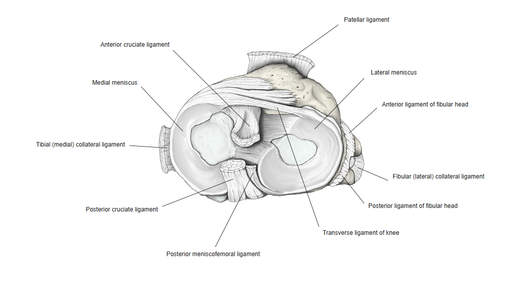
The medial meniscus is semicircular and is fused with the deep part of the tibial collateral ligament. Its two attachments lie relatively far apart, and it is wider behind than in front: the anterior horn is narrower than the posterior horn. Because of the way it is attached, it is much less mobile than the lateral meniscus. Because it is held so firmly, it cannot easily move out of the way: in lateral rotation of the leg it is strained and pulled hard (it is caught between the femur and tibia rather than gliding away), and in medial rotation it is relieved of load. This is why it is torn more often, especially when the bent knee is twisted.
The menisci move forwards and backwards as the knee bends and straightens; the lateral meniscus moves considerably more than the medial one. Their outer edge is joined to the joint capsule, and the thick outer part (a little under one third) is not pure cartilage and carries some blood vessels from the capsule. The thin inner part has no vessels: like the articular cartilage, which also has no vessels, it is nourished by the synovial fluid, has almost no power of self-repair, and does not heal by itself when it is torn, for example by overloading.
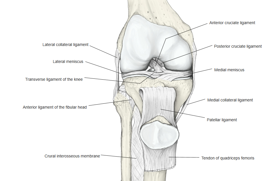
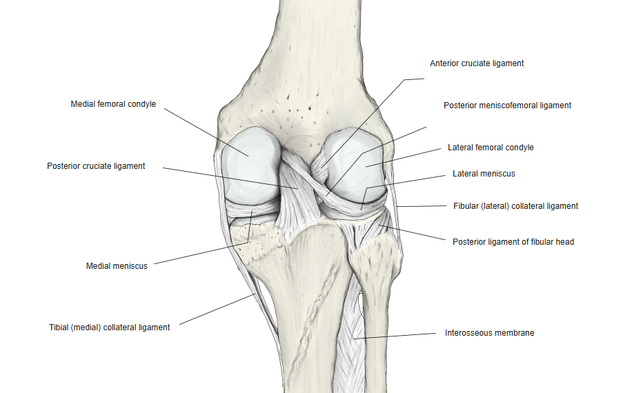
Note: the anterior horns of the two menisci are often joined by the transverse ligament of the knee. Some fibres of the semimembranosus reach the posterior part of the medial meniscus.
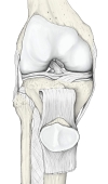 Right knee joint (anterior view)
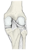 Right knee joint (posterior view)
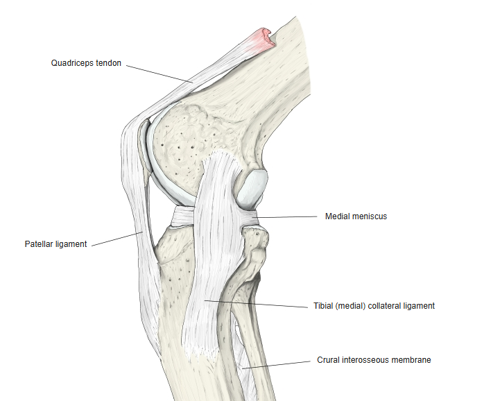
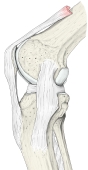 Right knee joint (medial view)
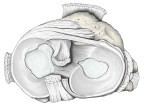 Menisci (superior view)
Reference websites
[Wikipedia]
· Medial meniscus, Wikipedia.
Related terms
- Lateral meniscus
- Articular meniscus
- Knee joint
- Medial condyle of tibia
- Medial condyle of femur
- Tibial collateral ligament
- Transverse ligament of knee
References and sources
- Further reading: Medial meniscus, Wikipedia.
- Federative International Programme for Anatomical Terminology (FIPAT). Terminologia Anatomica, 2nd ed. (TA2), 2019.