Nasolacrimal canal (Latin: canalis nasolacrimalis)
Also called the bony nasolacrimal canal
The nasolacrimal canal is the bony canal that runs from the floor of the orbit down to the nasal cavity and holds the nasolacrimal duct.
· It begins above at the fossa for the lacrimal sac at the medial corner of the orbit.
· Its walls are formed mainly by the maxilla (the lacrimal groove on its medial surface), completed medially by the lacrimal bone above and the lacrimal process of the inferior nasal concha below.
· It runs downward, slightly backward and laterally, and is about 12 mm long.
· It opens into the inferior nasal meatus, under the front part of the inferior nasal concha.
· It contains the membranous nasolacrimal duct, which carries tears from the lacrimal sac to the nose. The duct is often a little longer than the bony canal, so its opening may lie lower in the meatus.
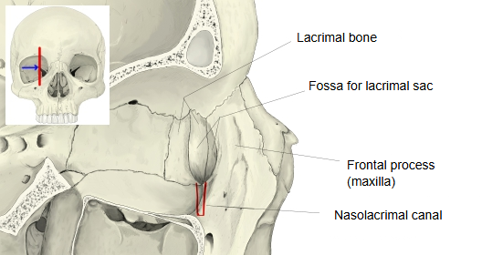
Where it is found
Related terms
- Fossa for lacrimal sac
- Lacrimal groove
- Inferior nasal meatus
- Maxilla
- Lacrimal bone
- Inferior nasal concha
- Nasolacrimal duct
References and sources
- Further reading: Nasolacrimal canal, Wikipedia.
- Further reading: Nasolacrimal duct, Wikipedia.
- Federative International Programme for Anatomical Terminology (FIPAT). Terminologia Anatomica, 2nd ed. (TA2), 2019.