Peyer's patches (Latin: noduli lymphoidei aggregati)
Also called Aggregated lymphoid nodules
- Overview
- Function
- Are the nodules of one patch independent?
- Reference websites
- (Wikipedia, websites with illustrations)
- Where it is found
- Related terms
Overview
· They are part of the immune system of the small intestine (the gut-associated lymphoid tissue) and start immune responses against microbes and other foreign material in the gut.
Distribution along the intestine
1. 1. Jejunum: few or almost none.
2. 2. Ileum: many, especially near its end; typical Peyer's patches are seen here.
3. 3. Large intestine: scattered single (solitary) lymphoid nodules are present, but they do not form grouped patches like Peyer's patches.
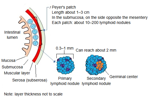
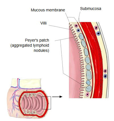
· One Peyer's patch contains about 10–200 primary and secondary lymphoid nodules (about 0.3–1 mm across each, growing to about 2 mm when active). The nodules sit in the mucosa and reach down into the submucosa; the villi over a patch are short or absent.
Function
1. 1. Taking up antigens
Specialised M cells in the covering epithelium take in microbes and particles from the gut and pass them to the immune cells of the lymphoid tissue beneath.
2. 2. Starting immune responses
B cells and T cells are activated; B cells mature into plasma cells that make IgA antibodies, which are secreted into the gut.
3. 3. Watching over the gut
They are a first line of defence against gut bacteria and pathogens entering through the intestinal wall.
Are the nodules of one patch independent?
Q: Is each lymphoid nodule of one Peyer's patch fully independent?
A: In short, the lymphoid nodules (follicles) of a Peyer's patch do not stand alone; they touch one another but are still distinct structural units — a "semi-independent" arrangement: separate, but not isolated.
In more detail:
1. Each follicle (lymphoid nodule) is an independent structural unit
Each has its own germinal centre
Follicular dendritic cells are present in each follicle
Each round to oval follicle is complete as a unit → in this sense they are independent lymphoid nodules.
2. But they are packed closely against their neighbours, and the borders are often not sharp.
In a Peyer's patch many follicles are crowded together
There is no wall (capsule) of connective tissue between the follicles
They lie in contact, and the connective tissue is continuous → in this sense they are not fully isolated.
3. The follicles are loosely separated by the interfollicular areas.
T-cell zones (parafollicular areas)
High endothelial venules (HEVs) are present
Differences in cell density in the interfollicular areas mark the borders between follicles
→ there is no wall, but there is a functional division.
Reference websites
[Wikipedia]
· Peyer's patch, Wikipedia.
[Websites with illustrations]
· Peyer's patches (images) (Wikimedia Commons)
· The small intestine (TeachMeAnatomy)
Where it is found
Related terms
References and sources
- Further reading: Peyer's patch, Wikipedia.
- Federative International Programme for Anatomical Terminology (FIPAT). Terminologia Anatomica, 2nd ed. (TA2), 2019.