Posterior arch of atlas (Latin: arcus posterior atlantis)
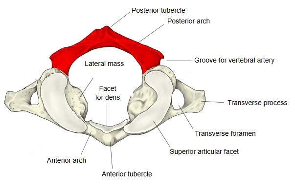
The posterior arch corresponds to the vertebral arch of other vertebrae and makes up about two-fifths of the ring; the shorter anterior arch closes the ring in front. In the midline it carries a small posterior tubercle, a rudimentary spinous process; the rectus capitis posterior minor arises from it and the nuchal ligament attaches to it. Its small size leaves room for movement between the atlas and the skull. On the upper surface, just behind each lateral mass, is the groove for the vertebral artery, which carries the vertebral artery and the suboccipital nerve; a thin bony bridge sometimes turns this groove into a foramen (arcuate foramen). Its upper border gives attachment to the posterior atlanto-occipital membrane; its lower border is joined to the axis by the posterior atlanto-axial membrane.
Reference websites
[Wikipedia]
· Atlas (anatomy): posterior arch, Wikipedia.
[Websites with illustrations]
· First cervical vertebra, or atlas (Gray's Anatomy, fig. 86) (Wikimedia Commons)

· Cervical spine: anatomy, ligaments, nerves and injury (Kenhub)
Related terms
- Atlas
- Lateral mass of atlas
- Axis
- Posterior atlanto-occipital membrane
- Rectus capitis posterior minor
- Vertebral artery
References and sources
- Further reading: Atlas (anatomy), Wikipedia.
- Federative International Programme for Anatomical Terminology (FIPAT). Terminologia Anatomica, 2nd ed. (TA2), 2019.