Pterygomandibular raphe (Latin: raphe pterygomandibularis)
Also called the pterygomandibular ligament
Its four sides:
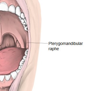
Medial surface: covered by the mucous membrane of the mouth. Here it raises a fold that can be seen when the mouth is opened wide.
Lateral surface: separated from the ramus of the mandible by adipose tissue (fat).
Posterior border: gives attachment to the superior pharyngeal constrictor.
Anterior border: gives attachment to the buccinator.
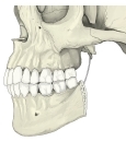 Pterygomandibular raphe (skull)
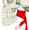 Pharyngeal constrictors
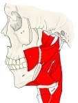 Buccinator and superior constrictor
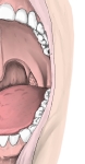 Open mouth
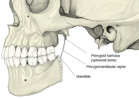
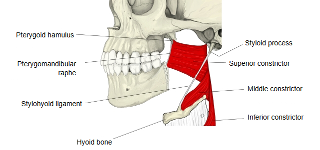
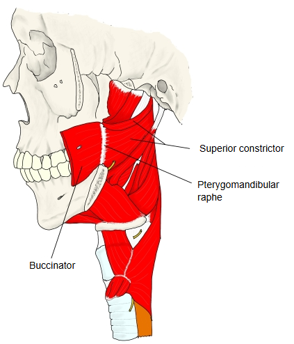
In short, it is a tendon between the pterygoid hamulus and the mandible that separates the buccinator from the pharyngeal constrictors.
It is a ligament-like band of connective tissue joining the pterygoid hamulus of the sphenoid bone to the back end of the buccinator crest of the mandible. It marks the boundary between the buccinator in front and the superior pharyngeal constrictor behind, but it is not always clearly seen. The fold it raises in the mouth is a landmark for the inferior alveolar nerve block.
Reference websites
[Wikipedia]
· Pterygomandibular raphe, Wikipedia.
· Buccinator muscle, Wikipedia.
· Superior pharyngeal constrictor muscle, Wikipedia.
[Websites with illustrations]
· Pterygomandibular raphe (Kenhub)
Sites with illustrations and photos


Related terms
- Pterygoid hamulus
- Buccinator
- Pharyngeal constrictors
- Medial plate of pterygoid process
- Mylohyoid line
- Retromolar triangle
- Pterygoid process
- Ramus of mandible
References and sources
- Further reading: Pterygomandibular raphe, Wikipedia.
- Federative International Programme for Anatomical Terminology (FIPAT). Terminologia Anatomica, 2nd ed. (TA2), 2019.