Superior ramus of ischium (Latin: ramus superior ossis ischii)
Also called the descending ramus of ischium
· Its back part forms the ischial tuberosity, which continues from the body.
· It has three surfaces: external, internal and posterior.
· The muscles and ligaments attached to it are those of the ischial tuberosity, together with the gemellus inferior and the obturator internus.
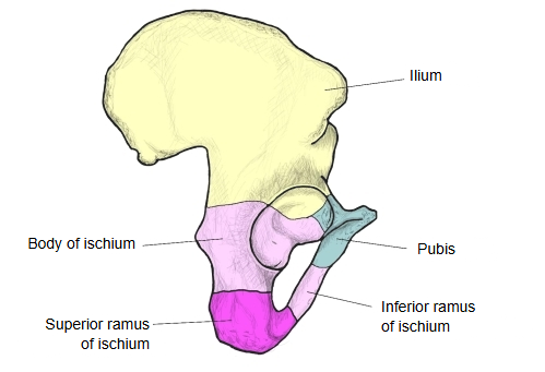
It is the part that continues on from the body below the acetabulum.
[Question]
Not all sources divide the ramus into superior and inferior parts: many describe the ischium only as a body and a ramus.
On that description the ischium forms the back and lower part of the hip bone and surrounds the obturator foramen from behind and below. The back and lower part of the acetabulum and the thick, three-sided part below it form the body; the thin, flattened bar running forwards, medially and upwards from the body is the ramus.
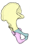 Parts of the hip bone
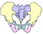 Bones of the pelvis
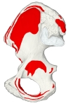 Muscle attachments (medial)
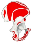 Muscle attachments (lateral)
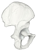 Hip bone (lateral view)
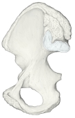 Hip bone (medial view)
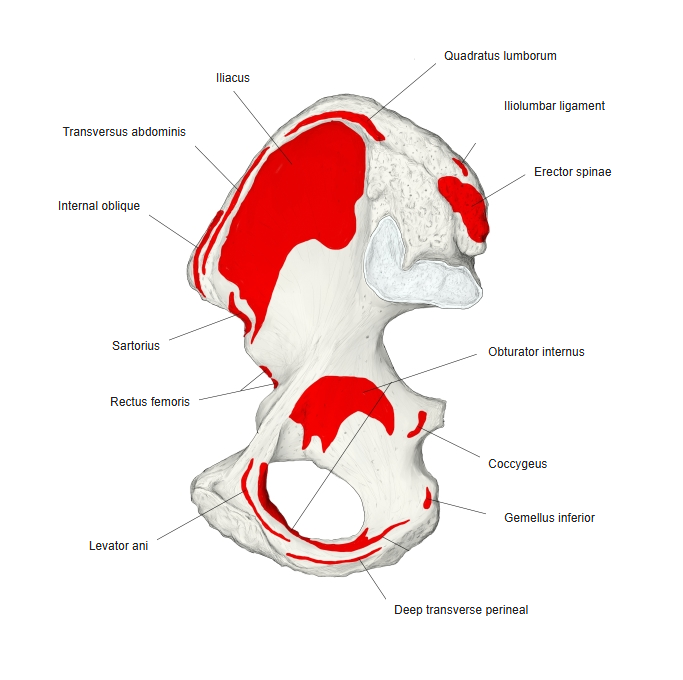
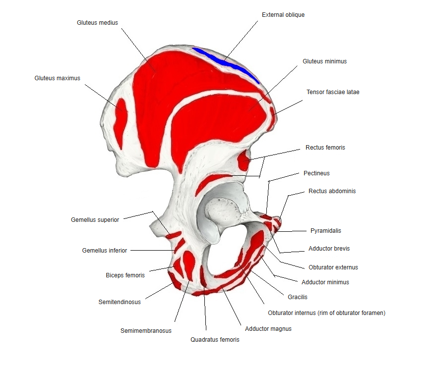
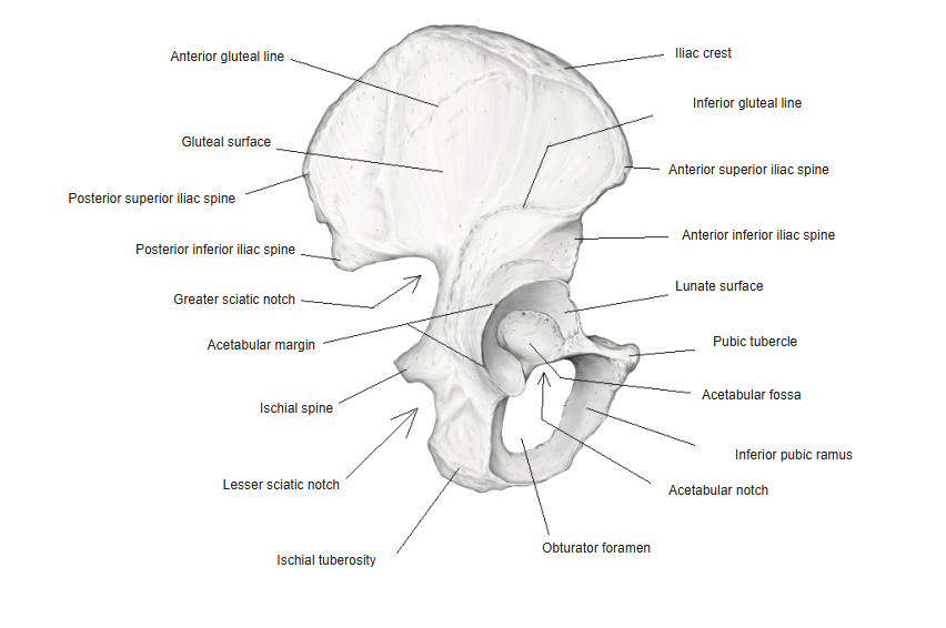
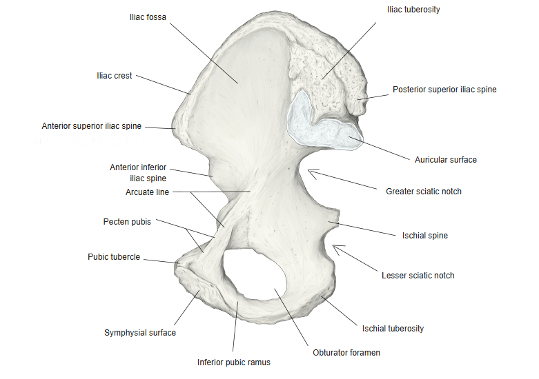
· The external surface is four-sided. A groove above it holds the tendon of the obturator externus; in front it is bounded by the posterior margin of the obturator foramen, and below it continues into the inferior ramus. It gives origin to the quadratus femoris, some fibres of the obturator externus and, lower down, part of the adductor magnus.
· The internal surface is part of the bony wall of the lesser pelvis. A sharp ridge at its lower edge receives the falciform process of the sacrotuberous ligament and, further forwards, gives origin to the superficial transverse perineal and ischiocavernosus muscles.
· Behind, the ramus forms the large swelling of the ischial tuberosity.
· Sources differ: Terminologia Anatomica divides the ischium only into a body (which includes the ischial tuberosity) and a ramus; the division of the ramus into superior and inferior rami is older usage (for example Gray's Anatomy, 1918). On that older division, the superior ramus is the lower part of the body in current terms, and the inferior ramus is the ramus.
Reference websites
[Wikipedia]
· Ischium, Wikipedia.
[Websites with illustrations]
· Right hip bone, external surface (Gray's Anatomy, fig. 235) (Wikimedia Commons)

· Right hip bone, internal surface (Gray's Anatomy, fig. 236) (Wikimedia Commons)

· Ischium: anatomy, structure and main parts (Kenhub)
Related terms
References and sources
- Further reading: Ischium, Wikipedia.
- Federative International Programme for Anatomical Terminology (FIPAT). Terminologia Anatomica, 2nd ed. (TA2), 2019.