Tarsal sinus (Latin: sinus tarsi)
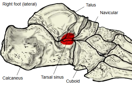
A funnel-shaped space formed by the calcaneal sulcus of the calcaneus and the sulcus tali of the talus; it is wide at its front lateral opening and narrows towards the back and the medial side. It opens on the lateral side of the foot just in front of the lateral malleolus; medially it narrows into the tarsal canal, which ends behind the sustentaculum tali. It separates the subtalar joint behind from the talocalcaneonavicular joint in front.
More details
The tarsal sinus contains the following.
| 1 | Blood vessels | Small arteries, including the artery of the tarsal canal (from the posterior tibial artery), which joins the artery of the tarsal sinus (from the arteries on the lateral side of the foot) to supply the talus. | |
| 2 | Nerves | Nerve endings, including sensory endings that are thought to help with the sense of the position of the foot. | |
| 3 | Ligaments | A ligament complex: · the medial, intermediate and lateral roots of the inferior extensor retinaculum · the cervical ligament · the interosseous talocalcaneal ligament (ligament of the tarsal canal) | 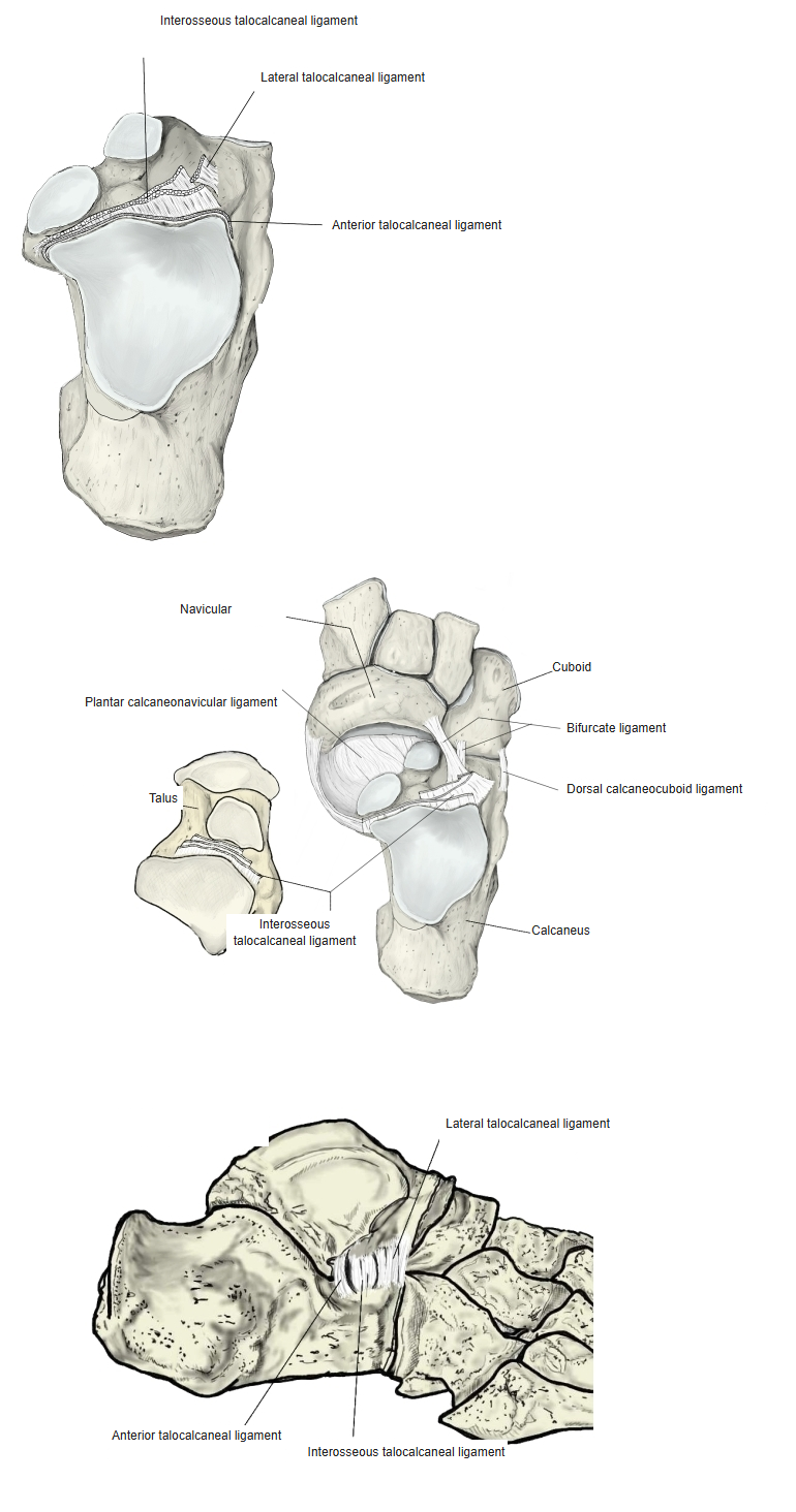 |
| 4 | Fat | Fatty tissue fills the rest of the space. |
Pain and tenderness over the lateral opening of the tarsal sinus, often after ankle sprains, is called sinus tarsi syndrome.
Reference websites
[Wikipedia]
· Sinus tarsi, Wikipedia.
· Subtalar joint, Wikipedia.
[Websites with illustrations]
· Bones of the foot: tarsals, metatarsals and phalanges (TeachMeAnatomy)
· Talus: anatomy and clinical aspects (Kenhub)
Where it is found
Related terms
- Calcaneal sulcus
- Sulcus tali
- Subtalar joint
- Talocalcaneonavicular joint
- Sustentaculum tali
- Talus
- Calcaneus
References and sources
- Further reading: Sinus tarsi, Wikipedia.
- Further reading: Subtalar joint, Wikipedia.
- Federative International Programme for Anatomical Terminology (FIPAT). Terminologia Anatomica, 2nd ed. (TA2), 2019.