Talocalcaneonavicular joint (Latin: articulatio talocalcaneonavicularis)
Also called the anterior lower ankle joint
Overview
- One of the intertarsal joints of the foot: a compound joint formed by several articular surfaces of the navicular, the talus and the calcaneus.
- The part between the talus and the navicular, together with the calcaneocuboid joint, makes up the transverse tarsal joint (or Chopart's joint).
Joints of the foot
- Joints of the tarsus : talocrural joint; intertarsal joints: talocalcaneonavicular, subtalar, transverse tarsal, calcaneocuboid, cuneonavicular, cuneocuboid, cuboideonavicular and intercuneiform joints
- Joints of the metatarsus : tarsometatarsal and intermetatarsal joints
- Joints of the toes : metatarsophalangeal and interphalangeal joints
Transverse tarsal joint (= Chopart's joint) = talocalcaneonavicular joint (the part between talus and navicular) + calcaneocuboid joint
The part between talus and navicular is also called the talonavicular joint.
Articular head and socket
| Articular surfaces | |
|---|---|
| Head | Articular surfaces of the head of the talus: navicular articular surface, anterior and middle calcaneal articular surfaces |
| Socket | In front: posterior articular surface of the navicular Medially and below: the fibrocartilage of the plantar calcaneonavicular ligament Below: anterior and middle talar articular surfaces of the calcaneus |
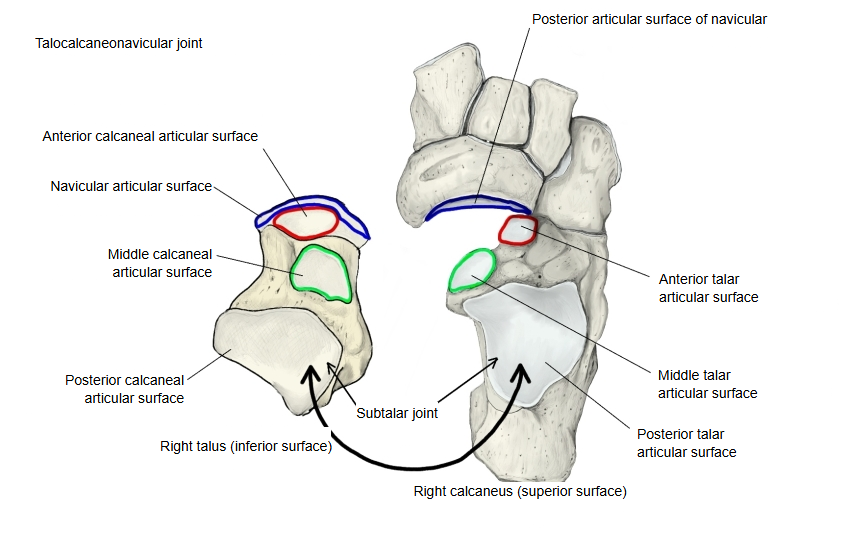
The plantar calcaneonavicular ligament stretches beneath the head of the talus and completes the socket. The joint has its own joint capsule.
The talocrural, subtalar and talocalcaneonavicular joints work together when the foot pushes off, as in jumping, and are therefore also called the ankle joints. The talocalcaneonavicular joint is the anterior lower ankle joint.
Ankle joints
- Upper ankle joint : ⇒ talocrural joint
- Lower ankle joint : anterior lower ankle joint ⇒ talocalcaneonavicular joint
posterior lower ankle joint ⇒ subtalar joint
Structure and movements
- The head and neck of the talus fit into a deep socket formed by the posterior articular surface of the navicular, the anterior and middle talar articular surfaces of the calcaneus, and the upper surface of the plantar calcaneonavicular ligament, which joins the calcaneus to the navicular.
- The joint cavity is separate and contains synovial folds with fat. The back of the capsule lies in the front part of the tarsal sinus and forms part of the interosseous talocalcaneal ligament.
- The dorsal wall of the capsule is the thin talonavicular ligament, running from the neck of the talus to the dorsal surface of the navicular; medially it blends with the tibionavicular part of the medial (deltoid) ligament.
- Together with the subtalar joint it turns the foot inward and outward about an axis that runs obliquely through the tarsus, from front-medial-upper to back-lateral-lower. In inversion the medial border of the foot is raised and the sole faces medially (comparable to supination of the hand); in eversion the lateral border is raised and the sole faces laterally (comparable to pronation).
- Combined with flexion and extension at the talocrural joint, these movements let the foot move around the talus, so that the tip of the foot can trace an oval.
- This joint and the calcaneocuboid joint together are called the transverse tarsal joint, or Chopart's joint.
Bones
Talocalcaneonavicular joint: navicular (posterior articular surface) ⇔ talus (navicular articular surface, anterior and middle calcaneal articular surfaces) ⇔ calcaneus (anterior and middle talar articular surfaces)
Type
Because the rounded head of the talus sits in a cup-shaped socket, many textbooks describe it as a ball-and-socket joint.
Ligaments
| Ligament | |
|---|---|
| 1 | Plantar calcaneonavicular ligament |
| 2 | Interosseous talocalcaneal ligament |
| 3 | Talonavicular ligament |
| 4 | Medial (deltoid) ligament |