Tympanosquamous fissure (Latin: fissura tympanosquamosa)
Also called the squamotympanic fissure
Between the upper edge of the front wall of the tympanic part and the back edge of the mandibular fossa there is a cleft that continues medially towards the sphenopetrosal fissure. In its lateral part it is a single fissure, the tympanosquamous fissure: the line of union of the squamous part and the tympanic part of the temporal bone.
The illustration below shows where the fissure lies on the lateral surface of the temporal bone.
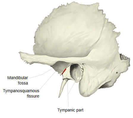
The front edge of the mandibular fossa rises gently to form the articular eminence (articular tubercle), to which the lateral ligament of the temporomandibular joint is attached. The back wall of the fossa is the thin bone of the tympanic part. Where this wall meets the fossa, the fissure is split medially by a thin edge of the petrous part (the lower edge of the tegmen tympani) into two lines: the petrosquamous fissure in front and the petrotympanic fissure behind. The chorda tympani (a branch of the facial nerve) and the anterior tympanic artery pass through the petrotympanic fissure and leave the skull there.
Reference websites
[Wikipedia]
· Petrotympanic fissure, Wikipedia.
· Temporal bone, Wikipedia.
[Websites with illustrations]
· Left temporal bone, outer surface (Gray's Anatomy) (Wikimedia Commons)

· Temporomandibular joint (Pocket Dentistry)
Related terms
- Tympanic part
- Squamous part
- Petrous part
- Tegmen tympani
- External acoustic meatus
- Chorda tympani
- Temporal bone
References and sources
- Further reading: Petrotympanic fissure, Wikipedia.
- Further reading: Temporal bone, Wikipedia.
- Federative International Programme for Anatomical Terminology (FIPAT). Terminologia Anatomica, 2nd ed. (TA2), 2019.